7 Pulmonary Arteries
D. Hortung, K. Hueper
Since anomalies of the pulmonary trunk or the pulmonary arteries are normally combined with heart defects or a patent ductus arteriosus (Botallo’s duct), they are more aptly classified as malformations. Many anomalies can be explained on the basis of embryological development of the aortic arches (Chapter 2). They can be classified as a systemic arterial blood supply to the lung, an atypical course of a pulmonary artery (sling formation), and the absence of one pulmonary artery.
The pulmonary arteries derive from the sixth aortic arch (Chapter 2). Whereas its distal part, the connection with the posterior aorta, disappears, it remains open on the left side until birth as the ductus arteriosus. Many anomalies can be attributed to the failure of an arch to develop normally or to a precipitous regression or a sudden hiatus of growth. 1–7
7.1 Normal Situation and Development

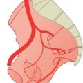
Fig. 7.1 Normal blood supply to the lung (>99%). Schematic (a) and contrast-enhanced CT (b). VR 3D image, oblique posterior view. 1 Left pulmonary artery; 2 aorta; 3 right pulmonary artery; 4 pulmonary trunk.

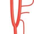
Fig. 7.2 Development of pulmonary arteries from sixth aortic arch. Schematic.
7.2 Supply by Systemic Arteries as Branches from the Aorta
An anomaly in which systemic arteries supply blood to the lung can be explained by the early stage of embryologie development during which branches of the posterior aortas are most often affected by this anomaly, which causes problems of higher systemic blood pressure and flow.2,8–12

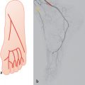
Fig. 7.3 Supply by systemic arteries as branches from the aorta (very rare). Schematic (a) and contrast-enhanced CT of the thoracic aorta from three patients (b–h). Patient 1: VR 3D images, posterior and lateral views (b), left lateral view (c), and MIP in sagittal view (d). Patient2: VR 3D images, posterior view and posterior view with descending aorta removed (e). Patient 3: VR 3D images, ventral and posterior views (f), right lateral oblique view (g), and MIP in transverse view (h). Major aortopulmonary collaterals are shown. 1 Pulmonary arteries; 2 descending aorta; 3 ascending aorta.
7.3 Sling Formation
Pulmonary slings are normally caused by an aberrant left pulmonary artery which arises from the right pulmonary artery and passes between the trachea and the esophagus to the left lung, producing respiratory distress. 13–18

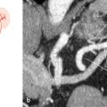
Fig. 7.4 Sling formation: aberrant left pulmonary artery from the right pulmonary artery passing between the trachea and the esophagus to the left (very rare). Schematic (a) and images from two patients (b–e). Patient 1: MRA, VR 3D images, lateral oblique view (b), frontal oblique view (c), and multi-planar reformation in transverse view (d). Patient 2: Contrast-enhanced CT; MIP in transverse view (e). 1 Left pulmonary artery; 2 right pulmonary artery; 3 trachea; 4 descending aorta.
Stay updated, free articles. Join our Telegram channel

Full access? Get Clinical Tree


