Procedure
Tolerance
Daily Tests
Mechanical
Laser localization
1 mm
Distance indicator (at isocenter)
2 mm
Collimator size indicator : both jaws and MLC
1 mm
Safety
Stereotactic interlocks (lockout): cone size, backup jaws
Functional
In addition to tests listed above (ASTRO)
Winston-Lutz test
≤0.75 mm average
IGRT positioning/repositioning
≤1 mm
Imaging subsystem interlocks
Functional
Monthly Tests
Dosimetry
Typical dose ratea output constancy
2 % (at SRS/SBRT dose rate, MU)
Mechanical
Treatment couch position indicatorsb
1 mm/0.5°
Localizing lasers
≤1 mm
In addition to tests listed above (ASTRO)
Winston-Lutz test: both cones and MLC, covering complete range of gantry, couch, collimator positions
≤0.75 mm average, ≤1 mm maximum
Hidden target test using SRS frame and/or IGRT system
≤1 mm
Annual Tests
Dosimetry
SRS arc rotation mode (range: 0.5–10 MU/deg)
Monitor units set vs delivered:
1 MU or 2 % (whichever is greater)
Gantry arc set vs delivered:
1° or 2 % (whichever is greater)
X-ray monitor unit linearity (output constancy)
±5 % (2–4 MU),
±2 % ≥ 5 MU
Mechanical
Coincidence of radiation and mechanical isocenter
±1 mm from baseline
Stereotactic accessories, lockouts, etc.
Functional
In addition to tests listed above (ASTRO)
Verification of small field beam data: output factors, depth dose, and off axis profiles for cones and MLC
±1 % from baseline
End-to-end localization assessment “hidden target test” using SRS frame and/or IGRT system
≤1 mm
End-to-end dosimetric evaluation using SRS frame and/or IGRT system
≤2 %
Table 5.2
SRS/SBRT-specific imaging-related quality assurance requirements
Procedure | Tolerance |
|---|---|
Daily Tests a | |
Planar kV and MV (EPID) imaging | |
Collision interlocks | Functional |
Positioning/repositioning | ≤1 mm |
Imaging and treatment coordinate coincidence (single gantry angle) | ≤1 mm |
Cone-beam CT (kV and MV) | |
Collision interlocks | Functional |
Positioning/repositioning | ≤1 mm |
Imaging and treatment coordinate coincidence | ≤1 mm |
Monthly Tests | |
Planar kV b and MV (EPID) imaging | |
Scalingc | ≤1 mm (kV), ≤2 mm (MV) |
Imaging and treatment coordinate coincidence (four cardinal angles) | ≤1 mm |
Spatial resolution, contrast, uniformity and noise | Baseline |
Cone-beam CT (kV and MV) | |
Geometric distortion | ≤1 mm |
Spatial resolution, contrast, HU constancy, uniformity and noise | Baseline |
Annual Tests | |
Planar kV imaging | |
Beam quality/energy | Baseline |
Imaging dose | Baseline |
Planar MV imaging (EPID) | |
Full range of travel SDD | ±5 mm |
Imaging dosed | Baseline |
Cone-beam CT (kV and MV) | |
Imaging dose | Baseline |
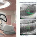
Table 5.1 presents SRS/SBRT-specific linac-related quality assurance requirements, which include the standard linear accelerator tests described in TG-142 and additional tests listed in “Quality and safety considerations in SRS and SBRT” in ASTRO [4]. Daily, monthly, and annual tests in Table 5.1 have specific recommendations based on the nature of the treatments delivered on the individual machine and are supplemented and modified as needed. Several tests deserve additional discussion. First, in addition to mechanical and safety tests in TG-142, a “Winston-Lutz” type of test provides the fundamental assessment of radiation isocenter and should be performed daily. The Winston-Lutz test can be performed by use of a film or an amorphous silicon electronic portal imaging device (EPID). Second, if image guidance is used for either cranial or extracranial localization, a test that verifies proper calibration and operation of those systems should also be performed daily. The Winston-Lutz test for both cones and MLC, covering complete range of gantry, couch, and collimator positions should be performed monthly. A hidden target test using SRS frame and/or IGRT system is performed monthly. Finally, end-to-end tests of both localization and dosimetric capabilities should be performed annually to assess the accuracy and integrity of the SRS/SBRT processes in an integrated manner.
Table 5.2 presents SRS/SBRT-specific imaging-related quality assurance requirements, which include the standard linear accelerator tests described in both the AAPM Task Group 142 report and ASTRO. The tests that verify geometrical arraignment, imaging quality, and imaging dose for planner kV and MV (EPID) imaging and cone-beam CT (kV and MV) should be performed appropriate frequency.
These SRS/SBRT-specific quality assurance requirements recommend annual evaluation of these characteristics. In addition, the report of AAPM task group 101 (TG-101) provides a number of excellent references for medical physicists, clinicians, and therapists in order to outline the best practice guidelines for SBRT [5]. The report of AAPM task group 135 will provide specific guidance for QA of robotic radiosurgery devices [6]. Similarly, the report of AAPM TG-148 describes QA for helical tomotherapy devices [7].
The TG-101 report includes a review of the literature to identify reported clinical findings and expected outcomes for the treatment modality. Information is provided for establishing a SBRT program, including protocols, equipment, resources, and QA procedures. Additionally, suggestions for developing consistent documentation for prescribing, reporting, and recording SBRT treatment delivery is provided. The major feature that separates SBRT from conventional radiation treatment is the delivery of large doses in a few fractions, which results in a high biological effective dose (BED). In order to minimize the normal tissue toxicity, conformation of high doses to the target and rapid fall-off doses away from the target is critical. The practice of SBRT therefore requires a high level of confidence in the accuracy of the entire treatment delivery process. In SBRT, confidence in this accuracy is accomplished by the integration of modern imaging, simulation, treatment planning, and delivery technologies into all phases of the treatment process; from treatment simulation and planning, and continuing throughout beam delivery.
In addition to these major features, there are other characteristics that distinguish SBRT from conventional radiation therapy (Table 5.3). These include a general increase in the number of beams used for treatment, the frequent use of noncoplanar beam arrangements, small or no beam margins for penumbra, and the use of inhomogeneous dose distributions and dose-painting techniques (including IMRT). All of these technology improvements result in the highly conformal dose distribution that characterizes the SBRT technique.
Table 5.3
Comparison of typical characteristics of 3D/IMRT radiotherapy and SBRT
Characteristic | 3D/IMRT | SBRT |
|---|---|---|
Dose/fraction | 1.8–3 Gy | 6–30 Gy |
No. of fractions | 10–30 | 1–5 |
CTV/PTV (gross disease + clinical extension): | GTV/CTV/ITV/PTV | |
Target definition | Tumor may not have a sharp boundary | (well-defined tumors: GTV = CTV) |
Margin | Centimeters | Millimeters |
Physics/dosimetry monitoring
Related posts:Stay updated, free articles. Join our Telegram channel
Full access? Get Clinical Tree
 Get Clinical Tree app for offline access

|