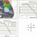
Fig. 7.1
An example of simple way of abdominal compression using bolus and body-shell
3.
Learning of regular respiratory patterns
Respiration with regular rhythm within fixed breathing range is important to achieve good result of RMC. Some kind of audio-feedback method such as utilization of metronome.
4.
Breath hold technique
Patient’s breath-hold is performed using some techniques. They include active breathing control [11], self-respiratory cessation with [12] or without [13] using respiratory indicator. Reproducibility of the breath-holding position is generally the best at the end of expiration [14], but has some differences among individuals.
5.
Gating with respiration
In conventional respiratory-gated radiotherapy, the gating window is usually set between the exhale baseline and a 20–30 % amplitude line and the irradiation beam will be triggered when part of the respiratory wave falls into this window. It was reported that respiratory internal margin could be reduced to 1.4 ± 0.7 mm by the gated technique [15].
6.
Tumor-tracking with respiration
It consists of two major aspects: real-time localization of, and real-time beam adaptation to, a constantly moving tumor. Compared to the breath-holding or gated method, tumor-tracking techniques potentially offer additional benefits such as higher delivery efficiency. The tumor-tracking is achieved with [16] or without [17] insertion of fiducial markers.
Respiratory status, timing of beam-on, time-efficiency, and general size of the internal margin according to the representative methods of RMC are shown in Fig. 7.2.
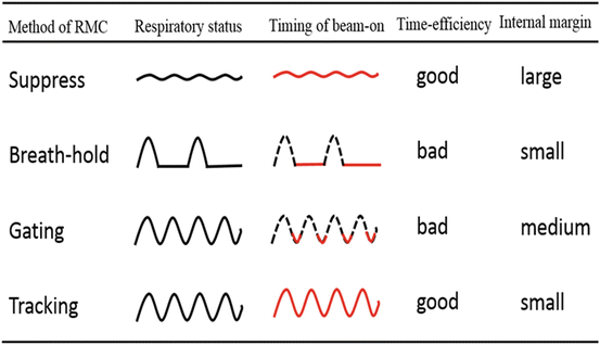
Fig. 7.2
Respiratory status, timing of beam-on, time-efficiency, and general size of the internal margin according to methods of respiratory motion control
7.5 Planning for the Treatment Under RMC
When performing RMC, a treatment plan must be established assuming the following uncertainties [2]: changes in the tumor form and organs at risk due to respiration; errors between the predicted and actual tumor positions, the length of time from sensing the respiratory phase to the actual initiation of irradiation. The CT examination for treatment planning session should be performed under the individual way according to the RMC method as shown Table 7.1. While fast scan CT or four-dimensional (4D) CT is recommended for the planning of the treatment under breath-hold, respiratory gated, or tracking technique, but fast scan CT is not tolerable for the planning of the treatment under shallow breathing. Slow-scan CT, generally taken with 4.0 or more seconds for a scanning of one-slice, is used for the planning of the treatment under shallow breathing in some facilities, however a particular attention should be paid for the use of slow-scan CT that may have deformity or blurring effect on the internal target volume [18].
Table 7.1
Recommended manners of CT examination for treatment planning session according to the each of RMC method
RMC methods | Planning CT |
|---|---|
Shallow breathing (Abdominal compression) | Four-dimensional (4D) or expiration and inspiration or (slow scan) |
Breath-holding | Fast scan |
Respiratory gating | Fast scan or 4D |
Tracking | Fast scan or 4D |
7.6 Cautionary Note in RMC
The most important preparation for a good RMC is a good understanding of the RMC technique and systems in each facility and instruction and exercise for the patient to understand well and perform regular and stabilized breathing through the treatment fraction.
Tumors often follow complex 3D trajectories and sometimes exhibit hysteresis [8]. Berbeco et al. have shown that, even if one models the tumor trajectory before the treatment, one projection image still may not localize tumor position with sufficient accuracy [19].
A baseline shift of respiratory status might be produced during a long period of SBRT or IMRT due to relaxation of the patient’s mental strain and the body-muscle.
Abdominal compression might cause the patient’s distress that can disturb the respiratory and body-wall condition.
As the tumor position under the same respiratory waveform pattern in a day does not always correspond to that in other days, confirmation of the tumor position using image-guidance is necessary in every treatment fraction.
When using fiducial markers, it should be noticed that comparative differences of the location between them and the tumor are changeable during whole treatment term because of migration of the markers or deformation of the lung.
The quality assurance and control (QA/QC) had better be done in every patient, radiotherapy fractions, and treatment methods or devices.
A guideline and manual of the RMC method in each facility must be made in combination with a good comprehension of all of radiotherapy staffs.
7.7 Devices for RMC
7.7.1 Measuring Instruments for Respiratory Motion
Common devices to verify the range of tumor motion before treatment are as follows.
X-ray fluoroscopy
CT at ends of inspiration and expiration
CT device is integrated with the therapeutic apparatus (cone-beam CT, megavolt CT, and CT-on-rails)
Four dimensional CT (4DCT)
Cine magnetic resonance imaging (MRI)
Cine electronic portal imaging device (EPID)
Common devices to verify the length of tumor motion during the irradiation are as follows.
Cine electronic portal imaging device (EPID)
X-ray fluoroscopy
Model which predicts the 3D position of a tumor from external breathing signals and others
Beacon transponder (Calypso, Varian Medical System Inc.)
7.7.2 Indicator for Respiratory Monitoring
Respiration status is essential to be monitored for all of RMC techniques. Most of the methods of respiratory monitoring are not according to lung volume itself but the change of body wall as a surrogate for respiratory status. The commonly-used on the market in Japan are as follows shown in Fig. 7.3.
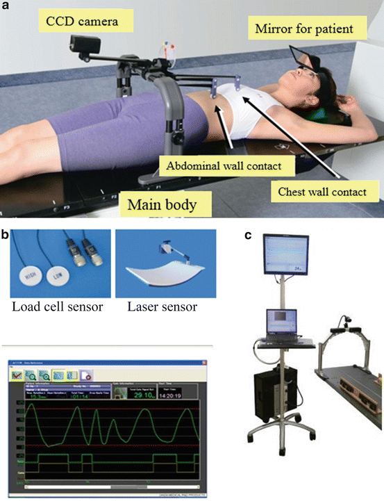
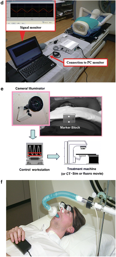
Fig. 7.3
(a) (Abches). (b) AZ-733 V(Anzai Medical Inc.). (c) Breath-track (Engineering System Inc.). (d) Air-bag system (Max Medical Inc.). (e) Real-time Position Management (RPM) system (Varian Medical Systems, Inc.). (f) Active breathing coordinator (ABC) system (Elekta Ltd.)
Air-bag system (Max Medical Inc. Fig. 7.3d)
Related posts:

Stay updated, free articles. Join our Telegram channel

Full access? Get Clinical Tree