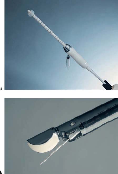
6 Radial EUS, Linear EUS, or Ultrasound Miniprobe: Which Is the Most Suitable Device in Gastrointestinal EUS? The Development of EUS: a Short Historical Survey Shortly after ultrasound first became available as a diagnostic imaging method in medicine,1 its use was expanded by Wild et al., who presented an intracavitary rectal application in 1957.2 The principle of using a blind, rigid probe has been maintained and further improved for vaginal and rectal ultrasonography (Fig. 6.1a). Endoscopic ultrasonography (EUS) with flexible instruments has developed quickly since the early 1980s3 and is now accepted as an indispensable tool in gastroenterology and associated fields. Apart from other indications, EUS is essential for the pretherapeutic local staging of gastrointestinal tumors. It is the method of choice for determining the origin and type of submucosal lesions. The use of longitudinal systems makes it possible to carry out fine-needle biopsy of lesions close to the gastrointestinal tract and local interventions guided by real-time EUS. Fig. 6.1a, b The modern rigid rectal probe (a) and flexible longitudinal echoendoscope (b) for diagnostic and interventional endoscopic ultrasonography. The development of flexible echoendoscopes started with mechanical radial scanners (5 MHz, 360°), which were mounted on top of a conventional endoscope.3 Soon after this, prototype echoendoscopes based on the radial mechanical technique were manufactured by the Olympus Corporation.4 Subsequent models worked with a frequency range of 5–12MHz and dominated clinical applications for some 10 years. Most of the relevant studies on diagnostic EUS were conducted using this type of instrument. At the beginning of the 1990s, a novel longitudinally scanning echoendoscope was presented and produced in series (Hitachi/Pentax FG-32UA).5 This instrument had an electronic scanner at frequencies of 5–10 MHz and allowed vessels to be examined using pulsed Doppler and color Doppler (Fig. 6.1b). In comparison with the mechanical devices, electronic scanners provided significantly better spatial resolution and higher imaging quality, especially in the near field. The fundamental advantage of the linear scanner is that it allows real-time controlled EUS-guided biopsy and interventions. Needles can be introduced through a working channel, and their tip is moved forward into the tissue within the scanning plane.6 The combination of these interventional instruments with color flow Doppler imaging reduces the risk of bleeding during puncture and transmural insertion of drains. Nowadays, modern radial scanners also work electronically, incorporating Doppler facilities and providing better spatial resolution. The ultrasound miniprobe was also developed at the same time as longitudinal scanners. The miniprobe is characterized by its small caliber and high ultrasound frequency (7.5–30 MHz), which provides high resolution, but at the expense of penetration depth. These probes were initially used in intracoronary ultrasonography,7–9 and the applications were later extended to the gastrointestinal field.10 The miniprobe is inserted into the working channel of a standard endoscope; endoscopy and EUS can thus be combined in a single examination. The small caliber of the instrument makes it possible to explore narrow tumor stenoses and allows intraductal biliary or pancreatic ultrasonography. Miniprobes usually work mechanically and produce a radial 360° image. The main disadvantage of these devices is their small penetration depth (2–3 cm) at frequencies of 20–30 MHz. Their use is therefore limited when the focus of the examination is on the surroundings of the gastrointestinal wall—e.g., for exploring lymph nodes and the pancreas (reviewed in11). The main obstacles to the widespread use of EUS are its limited (although still expanding) range of indications and the high initial costs and operating costs. It depends on the structure of local departments whether the purchase of one or more types of EUS instrument is economically justifiable. If only one system is to be bought, the choice of the correct type of EUS unit is also an issue. In centers with low annual numbers of examinations, it is economically mandatory for the basic ultrasound unit to be usable for transabdominal, vascular, and cardiac ultrasonography as well. All EUS equipment manufacturers now offer variable-use systems. The initial costs for a standard echoendoscope are about $ 60 000–90 000. Experience shows that the lifetime of an instrument used at a frequency of 500 examinations per year is ≈ 3 years. Purchasing a second echoendoscope is profitable above a frequency of three or four examinations per day. The lifetime of a miniprobe is highly dependent on the examiner’s level of experience and the care taken with the instrument, and on the uses to which it is put (e.g., esophageal applications versus intraductal ultrasonography). Reports on the average number of examinations conducted with one probe vary from 10 to 90. The price of a miniprobe is about $2000–4000. During the last 10 years, the quality of all of the EUS systems available on the market has significantly improved. However, there are specific differences between the systems. The following sections provide general comparisons between linear and radial standard EUS systems and ultrasound miniprobes, without concentrating on specific instruments that are currently available. Miniprobe Versus Conventional EUS The indisputable advantage of miniprobe ultrasonography is that it makes it possible to carry out endoscopy and EUS during a single session. An ideal indication for this method is the evaluation of submucosal tumors or unclear external impressions.11 The only precondition for this is that the basic ultrasound unit should be immediately available, rather than being used for routine examinations by another physician in a different room. High-frequency miniprobe ultrasonography is limited by its low penetration depth. Systematic exploration of the pancreas and lymph-node areas or other structures more distant from the gastrointestinal wall is therefore not generally possible, although it has been done successfully in several cases. The second disadvantage is that it is not possible to carry out real-time controlled fine-needle aspiration biopsy (FNAB) with the miniprobe.
Related posts:

Stay updated, free articles. Join our Telegram channel

Full access? Get Clinical Tree


