Radiation Biology


Radiation Biology


20.1 OVERVIEW

Rarely have beneficial applications and hazards to human health followed a major scientific discovery more rapidly than with the discovery of ionizing radiation. Soon after Roentgen’s discovery of x-rays in 1895 and Becquerel’s discovery of natural radioactivity in 1896, adverse biological effects from ionizing radiation were observed. Within months after their discovery, x-rays were being used in medical diagnosis and treatment. Unfortunately, the development and implementation of radiation protection techniques lagged behind the rapidly increasing use of radiation sources. Within the first 6 months of their use, several cases of erythema, dermatitis, and alopecia were reported among x-ray operators and their patients. Becquerel himself observed radiation-induced erythema on his abdomen from a vial of radium he carried in his vest pocket during a trip to London to present his discovery. Many years later, this effect was referred to as a “Becquerel burn.” The first report of a skin cancer ascribed to x-rays was in 1902, to be followed 8 years later by experimental confirmation. However, it was not until 1915 that the first radiation protection recommendations were made by the British Roentgen Society, followed by similar recommendations from the American Roentgen Ray Society in 1922.

The study of the action of ionizing radiation on healthy and diseased tissue is the scientific discipline known as radiation biology. Radiation biologists seek to understand the nature and sequence of events that occur following the absorption of energy from ionizing radiation, the biological consequences of any damage that results, and the mechanisms that enhance, compensate for, or repair the damage. A century of radiobiologic research has amassed more information about the effects of ionizing radiation on living systems than is known about almost any other physical or chemical agent.

This chapter reviews the consequences of ionizing radiation exposure, beginning with the chemical basis by which radiation damage is initiated and its subsequent effects on cells, tissues and organ systems, and the whole body. This is followed by a review of the concepts and risks associated with radiation-induced carcinogenesis, hereditary effects, and the special concerns regarding radiation exposure of the fetus.


20.2 DETERMINANTS AND CLASSIFICATION OF THE BIOLOGIC RESPONSE OF RADIATION


20.2.1 Determinants of Biologic Response

Many factors contribute to producing the overall biologic response to radiation exposure. At the highest level, these variables can be thought of as those associated with the radiation source and those of the biological system being irradiated. The identification of these biologic effects depends on the method of observation and the time following irradiation. Radiation-related factors include the absorbed dose (quantity) and dose rate as well as the type and energy (quality) of the radiation. The
radiosensitivity of a complex biologic system is determined by a number of variables. Some of these are inherent to the type of cells exposed while others relate to the cell’s current biochemical, mitotic, and oxygen tension status as well as many other variables at the time of irradiation. Damage observed at the molecular or cellular level may or may not result in clinically detectable adverse effects. Furthermore, although some responses to radiation exposure appear instantaneously or within minutes to hours, others take weeks, years, or even decades to appear.


20.2.2 Classification of Biologic Effects

Biologic effects of radiation exposure can be classified as either stochastic effects or tissue reactions (deterministic effects). A stochastic effect is one in which the probability of the effect occurring, rather than its severity, increases with dose. Radiation-induced cancer and hereditary effects are stochastic in nature. For example, the probability of radiation-induced leukemia is substantially greater after an exposure to 1 Gy than to 10 mGy, but there will be no difference in the severity of the disease if it occurs. Stochastic effects are believed not to have a dose threshold, because damage to a few cells or even a single cell could theoretically result in the production of the disease. Therefore, even minor exposures may carry some, albeit small, increased risk (i.e., increased probability of radiation-induced cancer or a genetic effect). It is this basic, but unproven, model that risk increases with dose and there is no threshold dose below which the magnitude of the risk goes to zero, that is the basis of modern radiation protection programs, a goal of which is to keep exposures as low as reasonably achievable (see Chapter 21). Stochastic effects are regarded as the principal health risk from low-dose radiation, including exposures of patients and staff to radiation from diagnostic imaging procedures.

If the radiation dose to tissue is very high, the predominant biologic effect is cell killing, which presents clinically as degenerative changes in the exposed tissue. In this case, the effects are classified as tissue reactions (previously called deterministic effects), for which the severity of the injury, rather than its probability of occurrence, increases with dose. Tissue reactions differ from stochastic effects in that they require much higher doses to produce clinically observable effects and there is a threshold dose below which the effect does not occur or is subclinical. Skin erythema, fibrosis, and hematopoietic system damage are some of the tissue reactions that can result from large radiation exposures. As there is substantial individual variability in response to radiation, the “threshold dose” is just an approximation of the dose that would result in the specified effect. Many of these effects are discussed in the sections entitled “Response of Organ Systems to Radiation” and “The Acute Radiation Syndrome.” Tissue reactions can be caused by severe radiation accidents and can be observed in healthy tissue that is unavoidably irradiated during radiation therapy. Although they have been observed following some lengthy, fluoroscopically guided interventional procedures (Koenig et al., 2001; Shope, 1996), they are unlikely to occur as a result of routine diagnostic imaging procedures or occupational exposure.


20.3 INTERACTION OF RADIATION WITH CELLS AND TISSUE

As discussed in Chapter 3, x- and γ-ray photon interactions in tissue, as well as radiations emitted during radionuclide decay, result in the production of energetic electrons. These electrons transfer their kinetic energy to surrounding matter via excitation, ionization, and thermal heating. Energy is deposited randomly and rapidly (in <10-8 seconds [s]), and the secondary ionizations set many more low-energy
electrons in motion, causing additional excitation and ionization along the path of the initial energetic electron. For example, a single 30 keV electron, set in motion following the photoelectric absorption of a single x-ray or γ-ray photon, can result in the production of over 1,000 low-energy secondary electrons (referred to as delta rays), each of which may cause additional excitation or ionization events in the tissue (Goodhead, 1994). This chain of ionizations ultimately gives rise to subexcitation electrons (i.e., electrons with kinetic energies less than the first excitation potential of liquid water, 7.4 eV) that become thermalized as they transfer their remaining kinetic energy by vibrational, rotational, and collisional energy exchanges with the water molecules. Observable effects such as chromosome breakage, cell death, oncogenic transformation, and acute radiation sickness, all have their origin in radiationinduced chemical changes in important biomolecules.


20.3.1 Low Energy Electrons and Complex Damage

The delta rays and other lower-energy electrons, set in motion following an initial ionizing event, produce a unique ionization pattern in which closely spaced ionizations occur over a very short range (˜4 to 12 nm) along the path of the primary ionization track. The energy deposition (˜100 eV) along the shorter tracks referred to as spurs, whose diameters are approximately 4 to 5 nm, result in an average of three ionizing events. It is estimated that 95% of the energy deposition events from x-rays and γ-rays occur in spurs (Hall and Giaccia, 2018). Longer and less frequent pearshaped tracks called blobs deposit more energy (˜300 to 500 eV) and thus on average result in more ionization events (˜12 ion pairs) over their path (˜12 nm). High concentrations of reactive chemical species (such as free radicals—discussed below) are produced in these spurs and blobs and they increase the probability of molecular damage at these locations (Fig. 20-1A and B). If ionizing events occur near the DNA, whose diameter (˜2 nm) is on the same order as that of these short ionization tracks, they can produce damage in multiple locations in the DNA in close proximity to one another. These lesions, initially referred to as locally multiply damaged sites, are more difficult for the cell to repair and may be repaired incorrectly, Figure 20-1C (Goodhead, 1994; Ward, 1988). Synonyms for this type of damage in common use today include clustered damage, complex damage, and multiply damaged sites (MDS).

While endogenous processes, such as oxidative metabolism, mainly produce isolated DNA lesions, the complex clustered damage, in which groups of several damaged nucleotides occur within one or two helical turns of the DNA, is a hallmark of ionizing radiation-induced DNA damage. However while the radiation-induced pattern of molecular damage is different from other oxidative events in the cell of endogenous and exogenous (e.g., chemotherapy) origin, many of the functional changes produced in molecules, cells, tissues, and organs cannot be distinguished from damage produced by these other sources.

Repair of radiation damage occurs at molecular, cellular, and tissue levels. A complex series of enzymes and cofactors repair most radiation-induced DNA lesions within hours and, to the extent possible, damaged cells are often replaced within days following irradiation. However, the clinical manifestation of radiation-induced damage may appear over a period of time that varies from minutes to weeks and even years depending on the radiation dose, cell type, and the nature and scope of the damage. Only a fraction of the radiation energy deposited brings about chemical changes; the vast majority of the energy is deposited as heat. The heat produced is of little biologic significance compared with the heat generated by normal metabolic processes. For example, it would take more than 4,000 Gy, a supralethal dose, to raise the temperature of tissue by 1°C.


FIGURE 20-1 A. Low-LET radiation like x-rays and γ-rays is considered sparsely ionizing on average; however, a majority of the radiation energy is deposited in small regions (on the scale of nanometers) via denser clusters of ionizations from low-energy secondary electrons. This illustration depicts primary and secondary electron tracks producing clusters of ionization events. The calculated number of tracks is based on a cell nucleus with a diameter of 8 µm. The track size is enlarged relative to the nucleus to illustrate the theoretical track structure. B. A segment of the electron track is illustrated utilizing a Monte Carlo simulation of clustered damage produced by ionizations and excitations along the path of a low-energy (300 eV) electron. Excitation and ionization along with secondary electrons are shown until the electron energy drops below the ionization potential of water (˜10 eV). C. DNA double helix drawn on the same scale as the ionization track. Complex clustered damage can result from closely spaced damage to the DNA sugar-phosphate backbone and bases from both direct ionizations and diffusion of OH radicals produced by the radiolysis of water molecules in close proximity (few nm) with the DNA (i.e., indirect effect). Multiple damaged sites are shown as green, or orange, explosion symbols that denote DNA strand breaks, or damaged bases, respectively. In this example, the result is a complex double-strand break, consisting of three strand breaks and three damaged bases, all within ten base pairs along the DNA. This type of complex DNA lesion is more difficult for the cell to repair and can lead to cell death, impaired cell function, or transformations with oncogenic potential. (Reprinted with permission from Goodhead DT. Energy deposition stochastics and track structure: what about the target? Radiat Prot Dosimetry. 2006;122(1-4):3-15. Copyright © Oxford University Press.)


20.3.2 Free Radical Formation and Interactions

Radiation interactions that produce biologic changes are classified as either direct or indirect. The change is said to be due to direct action if a biologic macromolecule such as DNA, RNA, or protein becomes ionized or excited by an ionizing particle or photon passing through or near it. Indirect action refers to effects that are the result of radiation interactions within the medium (e.g., cytoplasm) that create mobile, chemically reactive species that in turn interact with nearby macromolecules. Because approximately 70% of most cells in the body are composed of water, the majority (˜70%) of radiation-induced damage from medical irradiation is caused by radiation interactions with water molecules. The physical and chemical events that occur when radiation interacts with a water molecule lead to the formation of a number of different highly reactive chemical species. Initially, water molecules are ionized to form H2O+ and free electrons (e). The e rapidly thermalizes and becomes hydrated, with a sphere of water molecules orienting around the e to form a hydrated or aqueous electron (image). The image then reacts with another water molecule to form a negative water ion (H2O + image → H2O). These water ions are very unstable; each rapidly forms another ion and a free radical:

H2O+ + H2O → H3O+ + OH (Hydroxyl Radical)

H2O → OH + H• (Hydrogen Radical)


Free radicals are atomic or molecular species that have unpaired orbital electrons. They are denoted by a dot next to the chemical symbol of the element with the unpaired electron. Thus, free radicals can be radical ions (e.g., H2O+ and H2O), or electrically neutral (OH, H•). The hydrogen and hydroxyl radicals can be created by other reaction pathways, the most important of which is the radiation-induced excitation and disassociation of a water molecule (H2O* excitation → H• and OH). The H+ and OH ions do not typically produce significant biologic damage because of their extremely short lifetimes (˜10-10 s) and their tendency to recombine to form water. Free radicals are extremely reactive chemical species that can undergo a variety of chemical reactions. Free radicals can combine with other free radicals to form nonreactive chemical species such as water (e.g., H• + OH → H2O), in which case no biologic damage occurs, or with each other to form other molecules such as hydrogen peroxide (e.g., OH + OH → H2O2), which is toxic to the cell. However, for low linear energy transfer (LET) radiation like x- and γ-rays (see Chapter 3), the molecular yield of H2O2 is low and the majority of indirect effects are due to the interactions of the hydroxyl radicals with biologically important molecules.

The damaging effect of free radicals is enhanced by the presence of oxygen. Oxygen reacts with free radicals and reduces the probability of free radical recombination into nontoxic chemicals such as water or molecular hydrogen. Oxygen can combine with the hydrogen radical to form a highly reactive oxygen species (ROS) such as the hydroperoxyl radical (e.g., H• + O2 → HO2•). Free radicals can act as strong oxidizing or reducing agents by combining directly with macromolecules. Free radicals can attack biomolecules (R) in a number of ways, including hydrogen abstraction (RH + OH → • R• + H2O) and OH addition (R + OH → ROH•). Chemical repair, restitution, of the damaged biomolecules can occur via radical recombination (e.g., R• + H• → RH) or more commonly, by hydrogen donation from thiol compounds (RSH + R• → RH + RS•), producing the much less reactive or damaging thiyl radical. In the presence of oxygen, chemical repair is inhibited by the transformation of organic radicals into peroxyradicals (R• + O2 → RO2•). Because they are highly reactive, free radicals have limited lifetimes (less than 10-5 s) and very short diffusion distances, but they can diffuse sufficiently far in the cell (on the order of ˜4 nm) to produce damage at locations other than their origin. Free radical-induced damage to DNA is the primary cause of biologic damage from low-LET radiation. While radiation exposure from medical imaging does result in some direct ionization of critical cellular targets, approximately two thirds of the total radiation damage is due to the free radical-mediated indirect effects of ionizing radiation.

Many enzymatic repair mechanisms exist within cells that are capable, in most cases, of returning the DNA to its preirradiated state. For example, if a break occurs in a single strand of DNA, the site of the damage is identified and the break may be repaired by rejoining the broken ends. If the damage is more complex or too severe or the cell repair mechanisms are compromised or overwhelmed by excessive radiation exposure, the damage to the DNA could persist. The clinical consequence of such DNA damage depends on a number of variables. For example, if the damage were to the DNA at a location that prevented the cell from producing albumin, the clinical consequences would be insignificant considering the number of cells remaining with the ability to produce this serum protein. If, however, the damage were to the DNA at a location that was responsible for controlling the rate of cell division (e.g., in an oncogene or tumor suppressor gene), the clinical consequences could be the formation of a tumor or cancer. Heavily irradiated cells, however, often die during replication, thus preventing the propagation of seriously defective cells. Figure 20-2 summarizes the physical and biologic responses to ionizing radiation.


FIGURE 20-2 Physical and biologic responses to ionizing radiation. Ionizing radiation causes damage either directly by damaging the molecular target or indirectly by ionizing water, which in turn generates free radicals that attack molecular targets. The physical steps that lead to energy deposition and free radical formation occur within 10-5 to 10-6 s, whereas the biologic expression of the physical damage may occur from seconds to decades later.

Experiments with cells and animals have shown that the biologic effect of radiation depends not only on factors such as the dose, dose rate, environmental conditions at the time of irradiation, and radiosensitivity of the biologic system but also on the spatial distribution of the energy deposition at the molecular level (microdosimetry).


20.4 MOLECULAR AND CELLULAR RESPONSE TO RADIATION

Although all ionizing radiations are capable of producing similar types of biologic effects, the magnitude of the effect per unit dose differs. To evaluate the effectiveness of different types and energies of radiation and their associated LETs, experiments are performed that compare the dose required for the test radiation to produce the same specific biologic response produced by a particular dose of a reference radiation (typically, x-rays produced by a potential of 250 kV). The term relating the effectiveness of the test radiation to the reference radiation is called the relative biological effectiveness (RBE). The RBE is defined, for identical exposure conditions, as:


The RBE is initially proportional to LET. As the LET of the radiation increases, so does the RBE (Fig. 20-3). The increase is attributed to the higher specific ionization (i.e., ionization density) associated with high-LET radiation (e.g., alpha particles) and its relative advantage in producing cellular damage (increased number and
complexity of clustered DNA lesions) compared with low-LET radiation (e.g., x- and γ-rays). However, beyond approximately 100 keV/µm in tissue, the RBE decreases with increasing LET, because of the overkill effect. Overkill (or wasted dose) refers to the deposition of radiation energy in excess of that necessary to produce the maximal biologic effect. The RBE ranges from less than 1 to more than 20. For a particular type of radiation, the RBE depends on the biologic endpoint being studied. For example, chromosome aberrations, cataract formation, or acute lethality of test animals may be used as endpoints. Compared to high-energy γ-rays, the increased effectiveness of diagnostic x-rays in producing DNA damage is suggested not only by the differences in their microdosimetric energy deposition patterns but has also been demonstrated experimentally with an RBE of about 1.5 to 3. However, these differences do not necessarily imply (nor have epidemiological studies been able to confirm) an associated increase in cancer risk. The RBE also depends on the total dose, dose rate, fractionation, and cell type. Despite these limitations, the RBE is a useful radiobiologic tool that helps to characterize the potential damage from various types and energies of ionizing radiation. The RBE is an essential element in establishing the radiation weighting factors (wR) discussed in Chapter 3.


FIGURE 20-3 The RBE of a given radiation is an empirically derived value that, in general (with all other factors being held constant), increases with the LET of the radiation. However, beyond approximately 100 keV/µm, the radiation becomes less efficient due to overkill (i.e., the maximal potential damage has already been reached), and the increase in LET beyond this point results in wasted dose.

Although all critical lesions responsible for cell killing have not been identified, it has been established that the radiation-sensitive targets are located in the nucleus and not in the cytoplasm of the cell. Cells contain numerous macromolecules, only some of which are essential for cell survival. For example, there are many copies of various enzymes within a cell; the loss of one particular copy would not significantly affect the cell’s function or survival. However, if a key molecule, for which the cell has no replacement (e.g., DNA), is damaged or destroyed, the result may be cell death. In the context of diagnostic x-ray exposure, cell death does not mean the acute physical
destruction of the cell by radiation but rather a radiation-induced loss of mitotic capacity (i.e., reproductive death). There is considerable evidence that damage to DNA (Fig. 20-4) is the primary cause of radiation-induced cell death.


FIGURE 20-4 DNA is a primary target for damage that results in radiation-induced cell and tissue effects. The schematic illustrates the many orders of chromatin packaging from “naked” DNA to give rise to the highly condensed metaphase chromosome. The double helical DNA is wrapped around histones to form nucleosomes that are packaged to produce chromatin fibers that ultimately become highly packed in the chromosomes visible at mitosis. A mitotic chromosome is characterized by the centromere, which binds the two homologous chromatids together into the chromosome. The tips of each chromosome arm contain the telomeres. A gene is a sequence of nucleotides in a given position in the chromosome that is the functional unit of hereditary information, sometimes coding for a specific protein or controlling the function of other genetic material. (Courtesy of REAC/TS).


20.4.1 Radiation-Induced DNA Damage and Response


Spectrum of DNA Damage

The deposition of energy (directly or indirectly) by ionizing radiation induces chemical changes in large molecules that may then undergo a variety of structural changes. These structural changes include (1) hydrogen bond breakage, (2) molecular degradation or breakage, and (3) intermolecular and intramolecular cross-linking. The rupture of the hydrogen bonds that link base pairs in DNA may lead to irreversible changes in the secondary and tertiary structure of the molecule that compromise genetic replication and transcription. Molecular breakages also may involve the sugar-phosphate polymers that are the backbones of the two helical DNA strands. They may occur as single-strand breaks (SSBs), double-strand breaks (DSBs) (in which both strands of the double helix break simultaneously at approximately the same nucleotide pair), base loss, base changes, or cross-links between DNA strands or between DNA and proteins (Fig. 20-5B). An SSB between the sugar and
the phosphate can rejoin, provided there is no opportunity for the broken portion of the strands to separate. While the rejoining is not typically immediate, because the broken ends require the action of a series of enzymes (endonuclease, polymerase, ligase) to rejoin, the rejoining is fast and the repair typically occurs with high fidelity. The presence of oxygen potentiates the damage by causing peroxidation of a base, which then undergoes radical transfer to the sugar, causing damage that prevents rejoining.

A DSB can occur if two SSBs are juxtaposed or when a single, densely ionizing particle (e.g., an alpha particle) produces a break in both strands. DNA DSBs are very genotoxic lesions that can result in chromosome aberrations. The genomic instability resulting from persistent or incorrectly repaired DSBs can lead to carcinogenesis through activation of oncogenes, inactivation of tumor suppressor genes, or loss of heterozygosity. SSBs (caused in large part by the OH radical) are more easily repaired than DSBs and are more likely to result from the sparse ionization pattern that is characteristic of low-LET radiation. For mammalian cells, an absorbed dose of one Gy from x-rays will cripple the mitotic capability of approximately half of the cells exposed. Each cell would experience approximately 40 DSBs, 1,000 SSBs, and 3,000 damaged bases. While DSBs and complex DNA damage are often associated with high-LET radiation, in reality, all ionizing radiation is capable of producing a substantial number of complex DSBs. In the case of low-LET radiations, used in diagnostic imaging, about a quarter to a third of the absorbed dose in tissue is deposited via lowenergy secondary electrons with energies on the order of 0.1 to 5 keV. These lowenergy electrons produce high ionization densities over very short tracks that are of the same scale as the DNA double helix. The result is an increased probability of complex DNA damage that may contain not only SSBs and DSBs but localized base damage as well. These complex DNA lesions are less likely to be repaired correctly than an isolated SSB, DSB, or base damage, which may lead to permanent DNA modifications or losses (Goodhead, 1988, 1994). The higher effectiveness of alpha particles in producing biological damage, in comparison to low-LET radiation, is not due to an increased yield of DNA damage but rather the ability of the higher ionization density to produce more complex DNA lesions (Brenner and Ward, 1992) (Fig. 20-5A). The ability to produce several MDS in proximity in the chromatin structure is referred to as regional multiply damaged sites (RMDS). These lesions are repaired more slowly, if at all, and may serve as a signal for gene induction for a longer time than following low-LET irradiation (Löbrich et al., 1996). In addition, the production of RMDS increases the probability that short double-stranded oligonucleotides will be released, making high fidelity repair without the loss of sequence information problematic. Figure 20-5 illustrates some of the common forms of damage to DNA.

When one considers radiation damage to DNA, it is important to keep in mind that cellular DNA is not “naked,” but is highly organized, being wrapped around histones to form nucleosomes, which, in turn, are organized into chromatin fibers, which, ultimately, condense into the chromosomes that are visible at mitosis (Fig. 20-4). These increasing levels of complexity can also alter the radiation sensitivity of the DNA, as has been shown in studies that sequentially “simplified” the DNA structure from that in intact cells to isolated DNA.

Regardless of its severity or consequences, the loss or change of a base is considered a type of mutation. Although mutations can have serious implications, changes in the DNA are discrete and do not necessarily result in structural changes in the chromosomes. However, chromosome breaks produced by radiation do occur and can be observed microscopically during anaphase and metaphase, when the chromosomes are condensed. Radiation-induced chromosomal lesions can occur in both somatic
and germ cells and, if not repaired before DNA synthesis, may be transmitted during mitosis and meiosis. Chromosomal damage that occurs before DNA replication is referred to as chromosome aberrations, whereas that occurring after DNA synthesis is called chromatid aberrations. Unlike chromosomal aberrations, in chromatid aberrations, only one of the daughter cells will be affected if only one of the chromatids of a pair is damaged.


FIGURE 20-5 A. Ionization patterns for low- and high-LET radiations in DNA. B. Types of radiation-induced DNA damages. (Reprinted with permission from Japan Atomic Energy Agency, Dependence of Yield of DNA Damage Refractory to Enzymatic Repair on Ionization & Excitation of Density Radiation, 2007. Copyright © JAEA.)


DNA Repair

Repair of DNA damage from the decay of naturally occurring and internally incorporated radionuclides (primarily C-14 and K-40) occurs constantly. Because these and other elements (stable and radioactive forms) are in physiological equilibrium (being constantly replenished by ingestion and inhalation), there are approximately 7,000 atoms undergoing radioactive decay each second in the average person. The damage done to DNA by this radiation is repaired rapidly and with high fidelity. Even in the absence of radiation, DNA damage is a relatively common event in the life of a cell. Mammalian cells experience many thousands of DNA lesions per cell per day as a result of a number of common cellular functions such oxidative damage induced by ROS during metabolism and DNA synthesis (e.g., errors during base replication). It has been estimated that at least 10,000 oxidative DNA lesions are produced per cell per day by normal respiratory processes, and up to a million total DNA damages per cell per day by metabolic processes and environmental factors (Shrinivas et al., 2017). Yet the mutation rate is surprisingly low due to the effectiveness of the cell’s response and the varied and robust DNA repair mechanisms operating within the cell. DNA damage induces several cellular responses that enable the cell either to repair or to cope with the damage. For example, the cell may activate cell cycle checkpoints (which
arrest cell cycle progression) to allow for repair of damaged DNA or incompletely replicated chromosomes. In the case of potentially catastrophic DNA damage, the cell may initiate any of several cell death pathways (discussed below), effectively eliminating the damaged genetic material. The checkpoint and cell death responses (often known collectively as the DNA damage response [DDR]) utilize many of the same sensor molecules or complexes involved in DNA damage recognition and signal transduction. Many types of DNA repair mechanisms exist, including direct repair of a damaged nucleotide, base excision repair (BER), nucleotide excision repair (NER), SSB and DSB repair, and mismatch repair, each requiring its own set of enzymes (Fig. 20-6A). The repair of DNA damage depends on several factors, including the stage of the cell cycle and the type and location of the lesion.

There are specific endonucleases and exonucleases that, along with other proteins or complexes of proteins, are capable of repairing damage to the DNA. For example, most DNA base damage and SSBs are repaired by the BER pathway, involving enzymes that recognize the damage, enzymatic excision, use of the intact complementary DNA strand as the template on which to reconstruct the correct base sequence, and then a ligase to join DNA strands together (Fig. 20-6B). NER is the major pathway for the repair of bulky, helix-distorting lesions such as thymine dimers produced by exposure to ultraviolet radiation. While simple DNA lesions caused by metabolism and the actions of ROS are the primary substrates for the BER pathway, these lesions can also be recognized and repaired via the NER pathway. NER may play a more significant role in the repair of damage where significant distortions in the DNA structure occur. NER and BER processes are generally accurate and occur rapidly; approximately 90% of SSB and base damage are repaired within an hour after the initial damage. Even with DSBs, DNA rejoining is virtually complete within 24 hours (h) (Fig. 20-6C).

Since DNA DSBs (the simplest clustered lesion) are the most biologically important DNA damage caused by ionizing radiation, it is important to consider repair of DSBs in a bit more detail. DSBs can be repaired with high fidelity via homologous recombination repair (HRR) involving exchanges with homologous DNA strands (from sister chromatids after replication or from homologous chromosomes). More often DSBs are repaired by the error-prone nonhomologous end-joining (NHEJ) that involves end-to-end joining of broken strands (Fig. 20-7). Completely different sets of enzymes and repair processes are involved in HR and NHEJ (Fig. 20-8), and defects in a number of the enzymes, for example, ataxia telangiectasia mutated (ATM), components of the MRN complex, or ligase-4, result in increased radiosensitivity of individuals with those genetic defects.


Chromosomal Aberrations

HRR can preserve the genetic integrity of the chromosome while NHEJ repair results in loss of DNA fidelity. There is a strong force of cohesion between broken ends of chromatin material. Interchromosomal and intrachromosomal recombination may occur in a variety of ways, yielding many types of aberrations such as rings and dicentrics. Figure 20-9 illustrates some of the more common chromosomal aberrations and the consequences of replication and anaphasic separation. In some misrepair events, the two DSBs lead to a translocation (Fig. 20-9C). Translocations involve large scale rearrangements and can cause pre-carcinogenic alterations in cellular phenotype, but most do not impair cellular survival. Misrepair can also result in a dicentric chromosome aberration (Fig. 20-9D), which generally destroys the clonogenic viability of the cell.

The extent of the total genetic damage transmitted with chromosomal aberrations depends on a variety of factors, such as the cell type, the number and kind of genes deleted, and whether the lesion occurred in a somatic or in a gametic cell.


FIGURE 20-6 A. A summary of DNA repair processes. Each repair process is responsible for the repair of different types of DNA lesions. Some of the enzymes involved in each process are shown. Defects in some of these enzymes can lead to certain types of tumors or be targeted by certain drugs, as shown, for the treatment of cancers. (Reprinted with permission from Lord C, Ashworth A. The DNA damage response and cancer therapy. Nature. 2012;481:287-294. Copyright © Springer Nature.) B. Scientists have recently been able to visualize the complicated and dynamic structures of DNA ligase using a combination of x-ray crystallography and small-angle x-ray scattering techniques. These experiments revealed the crystal structure of the human DNA ligase I protein bound to a short DNA oligonucleotide. The ring-shaped structure in the center of the figure is the solvent-accessible surface of the protein. The extended, chromosomal DNA (long coils) is an artist’s representation of the high-level organization of DNA structure. The figure illustrates the ring-shaped ligase protein sliding along the DNA searching for a break in the phosphodiester backbone of the DNA that is the substrate for the enzyme’s DNA end-joining activity. The enzyme, DNA ligase, repairs millions of DNA breaks generated during the normal course of a cell’s life, for example, linking together the abundant DNA fragments formed during replication of the genetic material in dividing cells. DNA ligase switches from an open, extended shape to a closed, circular shape as it joins DNA strands together. (Courtesy of Tom Ellenberger, DVM, PhD, Department of Biochemistry and Molecular Biophysics at Washington University School of Medicine, St. Louis, MO.) C. DSB induction and repair in primary human fibroblasts. Using immunofluorescence techniques, a fluorescent antibody-specific for γ-H2AX (a phosphorylated histone) forms discrete nuclear foci that can be visualized at sites of DSBs. DSB repair was evaluated at 3 min and 24 h after exposure to 200 mGy; repair was almost complete at 24 h. The length of the white scale bar shown in the unirradiated control panel equals 10 µm. (Reprinted with permission from Rothkamm K, Löbrich M. Evidence for a lack of DNA double-strand break repair in human cells exposed to very low x-ray doses. Proc Natl Acad Sci U S A. 2003;100:5057-5062. Copyright © National Academy of Sciences.)


FIGURE 20-7 Comparison of the two pathways for repair of DNA double-strand breaks: homologous recombination (HR) and non-homologous end-joining (NHEJ). Because HR usually uses the sister chromatid for the repair, it can only occur in the late S or G2 phases of the cell cycle, after DNA replication, but it is a highly accurate repair. On the other hand, the more common repair process, NHEJ, can occur at any time in the cell cycle but is an error-prone process that ligates the broken ends of DNA together, often resulting in loss of genetic information.

Chromosomal aberrations are known to occur spontaneously. In certain circumstances, the scoring of chromosomal aberrations in human lymphocytes has been used as a biologic dosimeter to estimate the dose of radiation received after accidental exposure. Lymphocytes are cultured from a sample of the patient’s blood and then stimulated to divide, allowing a karyotype to be obtained. The cells are arrested at metaphase, and the frequency of rings and dicentrics are scored. Wholebody doses from penetrating radiation in excess of 250 mGy for acute exposure and
400 mGy for chronic exposure can be detected with confidence limits that do not include zero. Although many chromosomal aberrations are unstable and gradually lost from circulation, this assay is generally considered the most sensitive method for estimating recent exposure (i.e., within 6 months). More persistent, stable reciprocal translocations can be measured using fluorescence in situ hybridization (FISH). In this method, chromosomes are labeled with chromosome-specific fluorescent DNA probes, allowing translocations to be identified using fluorescent microscopy (Fig. 20-9E). Reciprocal translocations are believed to persist for a considerable period after the exposure, and this approach has been used as one of the methods to estimate the doses to survivors of the atomic bombs detonated in Hiroshima and Nagasaki decades ago.


FIGURE 20-8 Model of the key steps required for NHEJ and HR repair of DNA DSBs. (Reprinted with permission from Chowdhury D, Choi Y, Brault M. Charity begins at home: non-coding RNA functions in DNA repair. Nat Rev Mol Cell Biol. 2013;14:181-189. Copyright © Springer Nature.)


FIGURE 20-9 Examples of chromosomal aberrations and the effect of recombinations, replication, and anaphasic separation. A. A single break in one chromosome, which results in centric and acentric fragments. The acentric fragments are unable to attach to the mitotic spindle and are transmitted to only one of the daughter cells where they may remain in the cytoplasm. These fragments are eventually lost in subsequent divisions. B. Ring formation may result from two breaks in the same chromosome in which the two broken ends of the centric fragment recombine. The ring-shaped chromosome undergoes normal replication, and the two (ringshaped) sister chromatids separate normally at anaphase—unless the centric fragment twists before recombination, in which case the sister chromatids will be interlocked and unable to separate. C. Translocation may occur when two chromosomes break and the acentric fragment of one chromosome combines with the centric fragment of the other and vice versa, or (D) the two centric fragments recombine with each other at their broken ends, resulting in the production of a dicentric. E. Metaphase spread, containing a simple dicentric interchange between chromosomes 2 and 8 visualized with multiplex fluorescence in situ hybridization (mFISH). This technique utilizes fluorescently labeled DNA probes with markers specific to regions of particular chromosomes, which allows for the identification of each homologous chromosome pair by its own color. The color is computer generated based on differences in fluorescence wavelength among probes. (Reprinted with permission from Cornforth MN, et al. Chromosomes are predominantly located randomly with respect to each other in interphase human cells. J Cell Biol. 2002;159(2):237-244. Copyright © Rockefeller University Press.)


20.4.2 Response to Radiation at the Cellular Level

There are a number of potential responses at the cellular level following radiation exposure. Depending on a variety of inherent and conditional biologic variables related to the cell and its environment (e.g., cell type, oxygen tension, stage
in the cell cycle at the time of exposure) as well as a number of physical factors related to the radiation exposure (e.g., dose, dose rate, LET), a number of responses are possible such as delayed cell division, apoptosis, reproductive failure, genomic instability (delay expression of radiation damage), DNA mutations including phenotypic (including potentially oncogenic) transformations, bystander effects (damage to neighboring unirradiated cells), and adaptive responses (irradiated cells become more radioresistant). Many of these effects are discussed in more detail below. While a wide variety of the biologic responses to radiation have been identified, the study of radiation-induced reproductive failure (also referred to as clonogenic cell death or loss of reproductive integrity) is particularly useful in assessing the relative biologic impact of various types of radiation and exposure conditions. The use of reproductive integrity as a biologic effects marker is somewhat limited, however, in that it is applicable only to proliferating cell systems (e.g., stem cells). For differentiated cells that no longer have the capacity for cell division (e.g., muscle and nerve cells), cell death is often defined as loss of specific metabolic functions or functional capacity. One must also keep in mind that, with many of the assays described below, sensitivity to detect changes may be limited at low radiation doses and dose rates, so data obtained at higher doses are often back-extrapolated, using various mathematical models discussed below, to low doses and dose rates.


Cell Survival Curves

Cells grown in tissue culture that are lethally irradiated may fail to show evidence of morphologic changes for long periods; however, reproductive failure eventually occurs. The most direct method of evaluating the ability of a single cell to proliferate is to wait until enough cell divisions have occurred to form a visible colony. Counting the number of colonies that arise from a known number of individual cells irradiated in vitro and cultured provides a way to easily determine the relative radiosensitivity of particular cell lines, the effectiveness of different types of radiation, or the effect of various environmental conditions. The loss of the ability to form colonies as a function of radiation exposure can be described by cell survival curves.

Several mathematical models have been developed to describe the biological response to radiation. The shape of a cell survival curve reflects the relative radiosensitivity of the cell line and the random nature of energy deposition and subsequent biological effects. Survival curves are usually presented in graphical form, with the surviving fraction (SF) of cells plotted using a logarithmic scale on the y-axis, as a function of the radiation dose shown using a linear scale on the x-axis. In the multitarget model, the response to radiation is defined by three parameters: the extrapolation number (n), the quasithreshold dose (Dq), and the D0 dose (Fig. 20-10).

The D0 describes the radiosensitivity of the cell population under study. The D0 dose is the reciprocal of the slope of the linear portion of the survival curve, and it is the dose of radiation that produces, along the linear portion of the curve, a reduction to 37% in the number of viable cells. Radioresistant cells have a higher D0 than radiosensitive cells. A lower D0 implies less survival per dose. The D0 for mammalian cells ranges from approximately 1 to 2 Gy for low-LET radiation.

In the case of low-LET radiation, the survival curve of mammalian cells usually is characterized by an initial “shoulder” before the linear portion of the curve on the semilogarithmic plot. The extrapolation number, which gives a measure of the “shoulder,” is found by extrapolating the linear portion of the curve back to its intersection with the y-axis. The extrapolation number for mammalian cells ranges between 2 and 10; the larger the n, the larger the shoulder. Dq also defines the width of the shoulder region of the cell survival curve and is a measure of sublethal damage.
Sublethal damage is a concept based on experiments that show that when the radiation dose is split into two or more fractions, with sufficient time between fractions, the cell survival increases after low-LET radiation. The presence of the shoulder in a cell survival curve is taken to indicate that more than one ionizing event (“hit”), on average, is required to kill a cell and the reappearance of the shoulder when a large dose is delivered in fractions indicates that the cells are capable of repairing sublethal damage between fractions.


FIGURE 20-10 Typical cell survival curve illustrating the portions of the curve used to derive the extrapolation number (n), the quasithreshold dose (Dq), and the D0 dose.

The linear-quadratic (LQ) model is now the most often used to describe cell survival data where the SF is generally expressed as

SF (D) = eαDβD2

where D is the dose in Gy, α is the coefficient of cell killing that is proportional to dose (i.e., the initial linear component on a log-linear plot) and β is the coefficient of cell killing that is proportional to the square of the dose (i.e., the quadratic component of the survival curve). The two constants (α and β) can be determined for specific tissues and cancers to predict dose response. As described previously, cell killing (i.e., loss of clonogenic viability) occurs via misrepaired or unrepaired chromosome damage such as dicentric aberrations that are formed when pairs of nearby DSBs wrongly rejoin to one another. The double helix can undergo a DSB as the result of two different mechanisms: (1) both DNA strands are broken by the same radiation track (or “event”) and (2) each strand is broken independently, but the breaks are close enough in time and space to lead to a DSB. The linear (alpha) component of the survival curve represents the damage done by individual radiation particle tracks and is thus independent of dose rate. While the damage is partially repairable over time, α still represents the probability of cell death due to individual, noninteracting, particle tracks. This linear (single-hit kinetics) dose-response relationship dominates with high-LET radiation. The quadratic (beta) component of the survival curve represents the probability of cell death due to interactions between two or more individual particle tracks (i.e., dominates with low-LET radiation and follows multiple-hit kinetics) causing the curve to bend at higher doses and is sensitive to dose rate.
The LQ (or alpha-beta model, as many call it) is more commonly used than the previously described nD0 model, for several reasons: the LQ model is mechanistically based, it is more useful in radiotherapy for explaining fractionation effect differences between late responding normal tissues and early responding tissues or tumors, and the LQ model seems to fit most experimental data on human cell lines. The dose at which cell killing is equal from the linear (αD) and quadratic (βD2) contributions is referred to as the α/β ratio. The α/β ratio is a measure of the curvature of the cell survival curve and, thus, a measure of the sensitivity of different cell types to fractionation of radiation dose, Figure 20-11. For example, late responding normal tissues such as spinal cord or lung that have smaller α/β ratios of 3 or 4 are preferentially “spared” by fractionation compared to tumors and early responding normal tissues (gut, skin, bone marrow) where the α/β ratio is larger (8 to 12), indicating less ability to repair (i.e., more alpha component and less effect of fractionating the dose).


Modes of Radiation-Induced Cell Death

When irradiated cells fail to form a colony, it can be because of loss of proliferative capacity due to processes such as senescence, quiescence, or terminal differentiation, or because of cell death including mitotic catastrophe, apoptosis, autophagy, necroptosis, or necrosis, Most radiation-induced death in proliferating cells results from mitotic death/catastrophe that occurs when cells are unable to go through mitosis, generally because of chromosomal damage (discussed above). Those damaged cells may then exhibit demise by any of the other processes just mentioned. Nonproliferating cells may be lost through the regulated cell death processes of apoptosis, autophagy, or necroptosis, or unregulated necrosis. Each of those processes involves characteristic morphological changes in cells, as well as different pathways with distinct cascades of molecular events. Furthermore, cross-talk can occur among the different cell death pathways at various levels. Figure 20-12 diagrammatically compares morphological changes in cells undergoing apoptosis, autophagy, and necrosis. Figure 20-13 presents molecular cascades involved in apoptosis, necroptosis, and autophagy.

Apoptosis is a form of cell death that is characteristically different from cell necrosis in morphology and biochemistry, leading to the elimination of cells without releasing inflammatory substances into the surrounding area. Apoptosis results in cell shrinkage via nuclear condensation and extensive membrane blebbing, ultimately resulting in fragmentation of the cell into membrane-bound apoptotic
bodies composed of cytoplasm and tightly packed organelles that are eliminated by phagocytosis. Hallmarks of apoptosis include the sequential activation of caspases (cysteine-dependent aspartate-directed proteases) from pro-caspases; interactions of pro- and anti-apoptotic members of the bcl-2 family of proteins, many working at the level of the mitochondria; cleavage of multiple proteins; and, ultimately, cleavage of DNA between nucleosomes to form characteristic fragments consisting of multiples of the amount of DNA in a nucleosome. Extrinsic apoptosis is initiated at the cell surface with activation of death receptors such as CD95 (Fas) receptor, dimerization and activation of the initiator, or upstream caspase, caspase-8, which, in turn, activates the downstream caspase-3, which activates endonucleases and other proteases
to cleave DNA and many other cellular proteins. Intrinsic apoptosis is generally started at mitochondria where interactions of pro-apoptotic proteins, such as Bax and Bak, with anti-apoptotic Bcl-2 and Bcl-XL results in the release of cytochrome c from the mitochondria, formation of apoptosomes, activation of caspase-9, activation of caspase-3, and the cleavage of other cellular proteins and DNA. Radiation can induce apoptosis through DNA damage initiating the formation of pro-apoptotic proteins such as Noxa and Puma, which activate intrinsic apoptosis or upregulation of death receptors to begin extrinsic apoptosis pathways. This is an over-simplistic description of the processes, as there can be much cross-talk among pathways and regulation by other proteins, for example, p53 or XIAP (sex-linked inhibitor of apoptosis) at various steps.


FIGURE 20-11 The LQ model. The experimental data are fitted to a LQ function. There are two components to cell killing: One is proportional to dose (αD); the other is proportional to the square of the dose βD2. The dose at which the linear and quadratic components are equal is the ratio α/β. The LQ curve bends continuously but is a good fit to the experimental data for the first few decades of survival. (Reprinted with permission from Hall EJ, Giaccia AJ. Radiobiology for the Radiologist. 7th ed. Philadelphia, PA: Lippincott Williams and Wilkins; 2012.)


FIGURE 20-12 Comparison of morphological changes in cells undergoing three different modes of radiationinduced cell death—apoptosis, autophagy, and necrosis. A particular mode of cell death may predominate depending on cell type, radiation quality, and dose, and other environmental factors. (Reprinted with permission from Hotchkiss RS, Strasser A, McDunn JE, Swanson PE. Cell death. N Engl J Med. 2009;361(16):1570-1583.)


FIGURE 20-13 Cascades of molecular events involved in apoptosis (A), autophagy (B), and necroptosis (C). See text for explanations. (A and C: Reprinted with permission from Matt S, Hofmann TG. The DNA damageinduced cell death response: a roadmap to kill cancer cells. Cell Mol Life Sci. 2016;73:2829-2850. B: Reprinted from Hotchkiss RS, Strasser A, McDunn JE, Swanson PE. Cell death. N Engl J Med. 2009;361(16):1570-1583.)

Autophagy was initially recognized as a process by which cells that were starved of nutrients initiated a “self-digestion” of cellular components to obtain energy and thus promote survival. Autophagy can also remove damaged cellular molecules and components and can be activated by genotoxic stress, such as radiation-induced DNA damage. On the other hand, if cell damage is extensive, autophagy can result in cell death. As with apoptosis, at the molecular level autophagy involves complex sequences of protein changes and enzyme activations, with cross-talk among the cascades possible. Proteins of importance recognized in the autophagy cascades include Beclin, LC3, and a series of atg proteins. Ultimately, cells undergoing autophagy sequester the proteins or components to be digested into autophagosomes, which fuse with lysosomes containing the lytic enzymes. In cancer biology, autophagy can actually be a double-edged sword, in some cases removing excessively damaged cells or, in the case of therapy, being activated to kill cancer cells; on the other hand, if autophagy does not kill the cell, it may permit cell survival with damaged DNA to become, or allow continued growth of, a cancer cell.

Necroptosis, or regulated necrosis, is more recently recognized as a mode of programmed cell death, including genetically determined enzyme cascades in its molecular expression. Interestingly, necroptosis appears to involve protein ubiquitination steps, as well as the formation of a necroptosome, which includes several RIP (rest in peace) kinases. Although at this writing, necroptosis does not appear to be a large component of radiation-induced cell death, the relative importance of the various cell inactivation mechanisms, apoptosis, autophagy, senescence, etc. depends on many intrinsic and extrinsic factors including cell type, tissue environment, and radiation dose, dose rate, and type, to name a few.

When thinking about radiation-induced cell inactivation, it is also important to give consideration to the time between radiation exposure and the occurrence of the inactivation processes just described. Again, the picture is not simple, as it depends on cell type and environment as well as the cell inactivation mode. For example, the apoptosis process itself, as just described, occurs fairly rapidly, in many cases only requiring a half-hour or so from the first activation of upstream caspases to total cell demise into apoptotic bodies, but the time between initial damage induction, for example, DNA DSBs from irradiation, to the start of the apoptotic process is variable. In lymphocytes, in vitro, or in vivo, apoptosis is seen within hours after irradiation. However, in some cell types, for example, many cancer cells, the damaged cells and their progeny are capable of dividing as many as 4 or 5 times before apoptosis is initiated in many or all the progeny of the initially irradiated cells; this has been shown to require as long as a week. In another example, senescence, for example, in fibroblasts, may be activated immediately after irradiation, but the non-dividing cells may remain functional for up to weeks, before they may eventually be removed from the population by necrosis or apoptosis.


Factors Affecting Cellular Radiosensitivity

Cellular radiosensitivity can be influenced by a variety of factors that can enhance or diminish the response to radiation or alter the temporal relationship between the exposure and a given response. These factors can be classified as either conditional or inherent. Conditional radiosensitivities are those physical or chemical factors that exist before and/or at the time of irradiation. Some of the more important conditional factors affecting dose-response relationships are discussed in the following paragraphs, including dose rate, LET, and the presence of oxygen. Inherent radiosensitivity includes those biologic factors that are characteristics of the cells themselves, such as the mitotic rate, the degree of differentiation, and the stage in the cell cycle.


Conditional Factors

The rate at which a dose of low-LET radiation is delivered has been shown to affect the degree of biologic damage for a number of biologic endpoints including chromosomal aberrations, reproductive delay, and cell death. In general, high dose rates are more effective at producing biologic damage than low dose rates. The primary explanation for this effect is the diminished potential for repair of radiation damage. Cells have a greater opportunity to repair sublethal damage at low dose rates than at higher dose rates, reducing the amount of damage, and increasing the survival fraction. Figure 20-14 shows an example of the dose rate effect on cell survival.

Note that the broader shoulder associated with low-dose-rate exposure indicates its diminished effectiveness compared with the same dose delivered at a higher dose rate. This dose-rate effect is diminished or not seen with high-LET radiation primarily because the dense ionization tracks produce more complex, clustered DNA damage that cannot be repaired correctly. Therefore, for a given dose rate, high-LET radiation is considerably more effective in producing cell damage than low-LET radiation (Fig. 20-15).


FIGURE 20-14 Cell survival curves illustrating the effect of dose rate for low-LET radiation. Lethality is reduced because the repair of sublethal damage is enhanced when a given dose of radiation is delivered at a low versus a high dose rate.


FIGURE 20-15 Cell survival curves illustrating the greater damage produced by radiation with high-LET. At 10% survival, high-LET neutron radiation is three times as effective as the same dose of low-LET radiation in this example.

For a given radiation dose of low-LET radiation, a reduction in radiation damage is also observed when the dose is fractionated over a period of time. This technique is fundamental to the practice of radiation therapy. The intervals between doses (hours to a few days) allow the repair mechanisms in healthy tissue to gain an advantage over the tumor by repairing some of the sublethal damage. Figure 20-16 shows an idealized experiment in which a dose of 10 Gy is delivered either all at once or in five
fractions of 2 Gy with sufficient time between fractions for repair of sublethal damage. For low-LET radiation, the decreasing slope of the survival curve with decreasing dose rate (see Fig. 20-14) and the reoccurrence of the shoulder with fractionation (see Fig. 20-16) are clear evidence of repair.


FIGURE 20-16 Idealized fractionation experiment depicting the survival of a population of 106 cells as a function of dose. Curve A represents one fraction of 10 Gy. Curve F represents the same total dose as in curve A delivered in equal fractionated doses (D1 through D5) of 2 Gy each, with intervals between fractions sufficient to allow for repair of sublethal damage. (Modified from Hall EJ. Radiobiology for the Radiologist. 5th ed. Philadelphia, PA: Lippincott Williams & Wilkins; 2000.)

The presence of oxygen increases the damage caused by low-LET radiation by inhibiting the recombination of free radicals to form harmless chemical species and by inhibiting the chemical restitution of damage caused by free radicals. This effect is demonstrated in Figure 20-17, which shows a cell line irradiated under aerated and hypoxic conditions with low or high-LET radiations. The relative effectiveness of radiation to produce damage at various oxygen tensions is described by the oxygen enhancement ratio (OER). The OER is defined as the dose of radiation that produces a given biologic response in the absence of oxygen divided by the dose of radiation that produces the same biologic response in the presence of oxygen. Increasing the oxygen concentration at the time of irradiation has been shown to enhance the killing of otherwise hypoxic (and thus radioresistant) cells that can be found in some tumors. The OER for mammalian cells in cultures is typically between 2.5 and 3 for low-LET radiation. High-LET damage is not primarily mediated through free radical production, and therefore the OER for high-LET radiation can be as low as 1.0.


Inherent Factors

In 1906, two French scientists, J. Bergonie and L. Tribondeau, performed a series of experiments that evaluated the relative radiosensitivity of rodent germ cells at different stages of spermatogenesis. From these experiments, some of the fundamental characteristics of cells that affect their relative radiosensitivities were established. The law of Bergonie and Tribondeau states that radiosensitivity is greatest for those cells that (1) have a high mitotic rate, (2) have a long mitotic future, and (3) are undifferentiated. With only a few exceptions (e.g., lymphocytes), this law provides a reasonable characterization of the relative radiosensitivity of cells in vitro and in vivo. For example, the pluripotential stem/early progenitor cells in the bone marrow have a high mitotic rate, have a long mitotic future, are poorly differentiated, and are
extremely radiosensitive compared with other cells in the body. On the other end of the spectrum, the fixed postmitotic neurons found in the central nervous system (CNS) are relatively radioresistant (Fig. 20-18). This classification scheme was refined in 1968 by Rubin and Casarett, who defined five cell types according to characteristics that affect their radiosensitivity (Table 20-1).


FIGURE 20-17 Cell survival curves demonstrating the effect of oxygen during high (blue) and low (green) oxygen tension on the OER for high- and low-LET irradiation.

The stage of the cells in the reproductive cycle at the time of irradiation greatly affects their radiosensitivity. Figure 20-19 shows the phases of the cell reproductive cycle and several checkpoints that can arrest the cell cycle or interrupt the progression to allow for the integrity of key cellular functions to be evaluated and if necessary, repaired. Experiments indicate that, in general, cells exposed to low-LET radiation are most sensitive during mitosis (M phase) and the “gap” (G2) between S phase and mitosis, less sensitive during the preparatory period for DNA synthesis (G1), and least sensitive during late DNA synthesis (S phase). If one looks at full survival curves from cells irradiated during each cell cycle phase, it is clear that the sensitive M and G2 phase cells have straighter curves, that is, less repair of radiation damage, while the late S phase cells have a broader shoulder, more repair. Consistent with these differences in repair ability, cells irradiated with high-LET radiation show much less cell cycle phase dependence, as the survival curves for cells in all phases of the cycle have minimal shoulders.

In addition to variations in radiation sensitivity through the cell cycle, there are differences in radiation-induced cell cycle arrest. As described in the legend to Figure 20-19, cell cycle checkpoints are critical times at which the cell monitors its integrity to ensure that it should proceed to the next phase of the cycle. These processes are carefully orchestrated at the molecular level by the interplay of proteins called cyclins and cyclin-dependent kinases (cdks) and are controlled by regulatory proteins including inhibitors of cdks (INK4 and KIP family regulators and Cdk inhibitors). Radiation can alter the function of these proteins, and thus, alter cell cycle checkpoint activity. Best known is the radiation-induced cell cycle arrest in the G2
phase because when it is prevented by genetic alterations or drugs such as caffeine, cells are sensitized to radiation. Also important for radiation sensitivity is the ability to arrest in the G1 phase, which is highly dependent on cells having a functional p53 pathway (see bottom of Fig. 20-19). The tumor suppressor gene TP53 (so named because it encodes a phosphorylated protein with a molecular weight of 53 kDa) operates predominantly at the G1/S checkpoint. The p53 protein (discussed again in relation to radiation-induced carcinogenesis later in the chapter) induces cellcycle arrest through the up-regulation of cyclin-dependent kinase inhibitors and thus allows for repair of DNA damage. The CIP/KIP (CDK interacting protein/Kinase inhibitory protein) family is one of two families (CIP/KIP and INK4) of mammalian cyclin-dependent kinase (CDK) inhibitors (CKIs) involved in regulating the cell cycle. The CIP/KIP family members also have a number of CDK-independent roles involving regulation of transcription, apoptosis, and the control of the cell’s cytoskeleton. Thus the p53 protein activation of cyclin-dependent kinases can activate DNA repair mechanisms or, in the case of severe DNA damage, induce cell death via apoptosis.
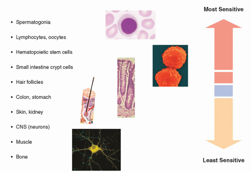


FIGURE 20-18 Relative radiosensitivity of tissues.


TABLE 20-1 CLASSIFICATION OF CELLULAR RADIOSENSITIVITY


CELL TYPE


CHARACTERISTICS


EXAMPLES


RADIOSENSITIVITY


VIM


Rapidly dividing; undifferentiated; do not differentiate between divisions


Type A spermatogonia


Erythroblasts


Crypt cells of intestines


Basal cells of the epidermis


Most radiosensitive


DIM


Actively dividing; more differentiated than VIMs; differentiate between divisions


Intermediate spermatogonia


Myelocytes


Relatively radiosensitive


MCT


Irregularly dividing; more differentiated than VIMs or DIMs


Endothelial cells


Fibroblasts


Intermediate in radiosensitivity


RPM


Do not normally divide but retain the capability of division; differentiated


Parenchymal cells of the liver and adrenal glands


Lymphocytesa


Bone


Muscle cells


Relatively radioresistant


FPM


Do not divide; differentiated


Some nerve cells


Erythrocytes


Spermatozoa


Most radioresistant


a Lymphocytes, although classified as relatively radioresistant by their characteristics, are in fact very radiosensitive. VIM, vegetative intermitotic cells; DIM, differentiating intermitotic cells; MCT, multipotential connective tissue cells; RPM, reverting postmitotic cells; FPM, fixed postmitotic cells.


Data from: Rubin P, Casarett GW. Clinical radiation pathology as applied to curative radiotherapy. Clin Pathol Radiat. 1968;22:767-768.


Adaptive Response, Bystander Effect, and Genomic Instability

A number of other responses to radiation have been observed in vitro that raise interesting questions about the applicability of the LQ dose-response model for lowdose low-LET radiation used in medical imaging. An adaptive response to radiation has been demonstrated in which an initial exposure or “priming dose” reduced the effectiveness of a subsequent exposure. For example, it has been demonstrated in vitro with human lymphocytes that compared to controls not receiving a small initial exposure, a priming dose of 10 mGy significantly reduced the frequency of chromosomal aberrations in the cells exposed to several Gy a few hours later (Shadley et al., 1987). However, the magnitude of this adaptive response varies considerably with

dose and dose rate as well as among lymphocytes from different individuals and with other variables. Many other endpoints for adaptive response have been studied such as cell lethality, mutations, and defects in embryonic development for which the evidence for an adaptive response was highly variable. While many theories have been advanced to explain this phenomenon, there is still insufficient evidence to use these results to modify the dose-response relationship for human exposure to radiation.


FIGURE 20-19 Phases of the cell’s reproductive cycle. The time between cell divisions is called interphase. Interphase includes the period after mitosis but before DNA synthesis (G1), which is the most variable in length of the phases; followed by S phase, during which DNA synthesis occurs; followed by G2, all leading up to mitosis (M phase), the events of which are differentiated into prophase, metaphase, anaphase, and telophase. Much of the control of the progression through the phases of a cell cycle is exerted by specific cell cycle control genes at checkpoints. Checkpoints are critical control points in the cell cycle that have built-in stop signals that halt the cell cycle until overridden by external chemical signals to proceed. There are three major checkpoints in the cell cycle, G1 checkpoint between G1 and S phase (G1/S), the G2 checkpoint between G2 and mitosis (G2/M), and the M (metaphase) checkpoint. The G1/S checkpoint is where the cell monitors its size, available nutrients, and the integrity of DNA (e.g., prevents copying of damaged bases, which would fix mutations in the genome) to assure all are adequate for DNA synthesis and progression on through the cell division cycle. In the absence of a proceed signal, the cell will enter a quiescent state G0 (the state of most cells in the body) until it receives a stimulation signal to continue. Following DNA synthesis, the G2/M checkpoint occurs at the end of the G2 phase, during which DNA damage induced during replication, such as mismatched bases and double-stranded breaks, is repaired. The cell ensures that DNA synthesis (S-phase) has been successfully completed before triggering the start of mitosis, and at the metaphase (spindle) checkpoint the cell monitors spindle formation and ensures all chromosomes are attached to the mitotic spindle by kinetochores prior to advancing to anaphase. p53, the product of the tumor suppressor gene TP53, operates predominantly at the G1/S checkpoint. The p53 pathway (inset) is vital to maintaining cell health, monitoring incoming stress from various sources such as oxidative stress, hypoxia, and DNA damage to name a few. Depending on the stressor, the response of the p53 pathway will change leading to cell cycle arrest, apoptosis, senescence, DNA repair, and metabolism adjustment.

The bystander effect is another fascinating phenomenon in which irradiated cells or tissues can produce alterations in nonirradiated cells or tissues. Sometimes also called the abscopal (“out-of-field”) effect of radiation, the effect has been demonstrated in a variety of experiments. One of the earliest examples was the ability of plasma from patients who had received radiation therapy to induce chromosomal aberrations in lymphocytes from nonirradiated patients (Hollowell and Littlefield, 1968). Among the most compelling evidence for the bystander effect are in vitro experiments in which an α-particle microbeam, with the ability to irradiate a single cell, can produce a host of changes in neighboring unirradiated cells. Examples of induced changes in the unirradiated cells include DNA damage such as micronuclei formation, sister-chromatid exchanges, cell killing, as well as changes in a number of important proteins (e.g., p53 and p21), genomic instability (discussed below), and even malignant transformation. Like adaptive response, the sequence of events following exposure is varied and complex and while many molecular mechanisms have been proposed to explain the bystander effects, the relationship of this phenomenon to low-dose, low-LET radiation effects characteristic of medical radiation exposure in humans is still an open question.

While the vast majority of unrepaired and misrepaired radiation-induced lesions are expressed as chromosomal damage at the first division, a fraction of cells can express chromosomal damage such as chromosomal rearrangements, chromatid breaks and gaps, and micronuclei over many cell cycles after they are irradiated. The biological significance and molecular mechanism surrounding this persistent genomic instability have been an area of active research for many years. Genomic instability has been demonstrated in vitro as delayed lethality in which cell cloning efficiency is reduced several generations after irradiation. Another interesting aspect is the differences in the types of mutations associated with radiation-induced genomic instability. Experiments have shown that the unirradiated progeny of the irradiated cells primarily demonstrate a de novo increase in lethal point mutations several generations after the initial irradiation. These mutations are more typical of spontaneous mutations than deletions and other mutations induced directly by ionizing radiation. There is evidence that suggests that errors induced during DNA repair may contribute to genomic instability. For example, experiments with cells deficient in the repair enzymes needed for NHEJ repair of radiation-induced DSBs demonstrate greater genomic instability than normal cells of the same type (Little, 2003). However, there are data to suggest that many other factors such as ROS, alterations in signal transduction pathways, centrosome defects, and other factors also play a role in radiation-induced genomic instability. Despite the many experimental models that have revealed different aspects of this phenomenon, the search for the relevance of radiation-induced genomic instability to radiation-induced cancer continues.

While radiation-induced responses such as genomic instability, adaptation, bystander effects, (as well as others which have not been discussed such as low-dose hypersensitivity) are fascinating in their own right, the results obtained are often restricted to specific experimental conditions and clear mechanistic understanding about these phenomena is still lacking. It has been suggested that these effects may alter the responses of cells and tissues to low doses of radiation, especially for carcinogenesis induction; however, at this time, they cannot be used reliably as modifying
factors to predict the biological consequences of radiation exposure in humans. On the other hand, the nonlinear nature of these and other multicellular and tissue-level responses raises serious questions regarding the current paradigm of linear extrapolation of risk based on the individual cell and the target. An active area of current research focused on addressing these complex responses to radiation interactions is a multidimensional, systems-level approach that includes the integration of radiation epidemiology with radiobiological investigations.


20.5 TISSUE AND ORGAN SYSTEM RESPONSE TO RADIATION

The response of tissues and organ systems to radiation depends not only on the dose, dose rate, and LET of the radiation but also on the relative radiosensitivities of the cells that comprise both the functional parenchyma and the supportive stroma. In this case, the response is measured in terms of morphologic and functional changes of the tissues and organ systems as a whole rather than simply changes in cell survival and kinetics.

The response of an organ system after irradiation occurs over a period of time. The higher the dose, the shorter the interval before the physiologic manifestations of the damage become apparent (latent period), and the shorter the period of expression during which the full extent of the radiation-induced damage is evidenced. There are practical threshold doses below which no clinically significant changes are apparent. In most cases, the pathology induced by radiation is indistinguishable from pathology caused by other physical, chemical, or biological agents or (in some cases) even naturally occurring diseases of unknown etiology.


20.5.1 Characterization of Radiosensitivity

Classically, the characterization of a tissue or organ system as radioresistant or radiosensitive was thought to depend in large part on the radiosensitivity of cells that comprised the functional parenchyma, with cells of the supportive stromal tissue consisting mainly of cells of intermediate radiosensitivity. Therefore, when the parenchyma contains radiosensitive cell types (e.g., stem/early progenitor cells in bone marrow or GI tract), the initial hypoplasia and concomitant decrease in functional integrity will be the result of damage to these radiosensitive cell populations, and functional changes are typically apparent within days or weeks after the exposure. However, if the parenchyma is populated by radioresistant cell types (e.g., nerve or muscle cells), it was thought that damage to the functional layer occurred indirectly by compromise of the cells in the vascular stroma, and hypoplasia of the parenchymal cells is typically delayed several months. The relative radiosensitivity of various tissues and organs and the primary mechanism for radiation-induced parenchymal hypoplasia, as described initially by Rubin and Casarrett, is shown in Table 20-2.

However, it is now clear that cell killing alone cannot explain many tissue reactions (ICRP, 2012), as those reactions also depend on complex events including inflammatory, chronic oxidative, and immune reactions, as well as damage to the vasculature and the extracellular matrix (ECM). In general, early reactions, such as in skin and GI tract, involve killing of the stem/early progenitor cells that supply the mature functional cells in the tissue, as well as inflammatory reactions. On the other hand, late reactions, for example, in the lung, kidneys, and brain, involve complex and dynamic interactions between multiple cell types in the tissues and organs and include infiltrating immune cells, production of cytokines and growth factors, often in persistent, cyclic cascades, and chronic oxidative stress. Cytokines are a diverse
group of soluble short-acting proteins, glycoproteins, and peptides produced by various immune and vascular cells that activate specific receptors and modulate the functions of many cells and tissues. Some cytokines may be membrane-bound or associated with ECM. Cytokines released by the vascular endothelium of irradiated tissues are implicated in the acute phase response to ionizing radiation and other inflammatory stimuli. Examples of radiation-induced cytokines include tumor necrosis factor (TNF3)-α, Interleukin (IL)-1, transforming growth factor (TGF)-β, and stem cell factor. Although many of these cytokines and growth factors, when induced by radiation, increase tissue damage, for example, the role of TGF-β in pneumonitis
is well documented, some growth factors can be radioprotective in some tissues, e.g., basic fibroblast growth factor protects microvasculature and IL-1 is a radioprotector of hematopoietic cells (Hall and Giaccia, 2018).


TABLE 20-2 RUBIN AND CASARETT CLASSIFICATION OF RELATIVE ORGAN AND TISSUE RADIOSENSITIVITY AND PRIMARY MECHANISM FOR RADIATION-INDUCED PARENCHYMAL HYPOPLASIA


ORGANS


RELATIVE RADIOSENSITIVITY


CHIEF MECHANISM OF PARENCHYMAL HYPOPLASIA


Lymphoid organs; bone marrow; testes and ovaries; small intestines


High


Destruction of parenchymal cells, especially the vegetative and differentiating cells


Skin and other organs with epithelial cell lining (cornea, lens, oral cavity, esophagus, GI organs, bladder, vagina, uterine cervix, uterus, rectum)


Fairly high


Destruction of radiosensitive vegetative and differentiating parenchymal cells of the epithelial lining


Growing cartilage; the vasculature; growing bones


Medium


Destruction of proliferating chondroblasts or osteoblasts; damage to the endothelium; destruction of connective tissue cells and chondroblasts or osteoblasts


Mature cartilage or bone; lungs; kidneys; liver; pancreas; adrenal gland; pituitary gland; thyroid; salivary glands


Fairly low


Hypoplasia secondary to damage to the fine vasculature and connective tissue elements


Muscle; brain; spinal cord


Low


Hypoplasia secondary to damage to the fine vasculature and connective tissue elements, with little contribution by the direct effect on parenchymal tissues


Note: Cells of the testes are more sensitive than ovaries. Skin radiosensitivity is particularly high around the hair follicles.


Adapted with permission from Rubin P, Casarett GW. Clinical radiation pathology as applied to curative radiotherapy. Clin Pathol Radiat. 1968;22:767-768.


FIGURE 20-20 Histopathology showing radiation-induced arteriole fibrosis (left). (From Zaharia M, Goans RE, Berger ME, et al. Industrial radiography accident at the Yanango hydroelectric power plant. In: Ricks RC, et al., eds. The Medical Basis for Radiation Accident Preparedness, the Clinical Care of Victims. New York, NY: The Parthenon Publishing Group; 2001:267-281.)

Important components of late responses also reflect damage to vasculature and development of fibrosis, caused by premature senescence and accelerated postmitotic differentiation leading to excessive collagen production by mesenchymal cells such as fibroblasts. Late radiation effects on the vasculature include fibrosis, proliferation of myointimal cells, and hyaline sclerosis of arterioles, the effect of which is a gradual narrowing of the vessels and reduction in the blood supply to the point that the flow of oxygen and nutrients is insufficient to sustain the cells comprising the functional parenchyma (Fig. 20-20). Importantly, these multiple interactions of the elements of tissue reactions change and evolve over time.


20.5.2 Healing

Healing of tissue damage produced by radiation occurs by means of cellular regeneration (repopulation) and replacement (Fig. 20-21). Regeneration refers to repopulation of the damaged cells in the organ by cells of the same type, thus recovering the lost functional capacity. Replacement refers to the development of fibrotic scar tissue, in which case the functionality of the organ system is compromised. The types of response and the degree to which they occur are functions of the dose, the volume of tissue irradiated, and the relative radiosensitivity and regenerative capacity of the cells that comprise the organ system. In so far as repopulation at the cellular level occurs within days after irradiation (Trott, 1991), fractionation of the dose (e.g., multiple fluoroscopically guided interventional procedures separated by days or weeks) allows for cellular repair and cellular repopulation and typically results in less extensive tissue damage than if the same total dose were to be delivered all at once. If the exposures are excessive, the ability of the cells to affect any type of healing may be lost, resulting in tissue fibrosis and necrosis.


FIGURE 20-21 Schematic diagram of organ system response to radiation.


20.5.3 Specific Organ System Responses

This section focuses on radiation-induced changes to the skin, reproductive organs, and eyes. Effects on the hematopoietic, gastrointestinal, and cardiovascular systems and the CNS are addressed in the context of the acute radiation syndrome (ARS). Additional information on radiation effects on these and other tissues and organ systems can be found in several textbooks and review publications listed at the end of the chapter under sections for suggested reading and references (e.g., Hall and Giaccia, 2018; ICRP, 2011; Mettler and Upton, 2008).


Skin

While radiation-induced skin damage is a relatively rare event, it is still the most commonly encountered tissue reaction (deterministic effect) following high-dose fluoroscopically guided interventional procedures. Acute radiation-induced skin changes were recognized soon after the discovery of x-rays and were reported in the literature as early as 1896 (Codman, 1902; Daniel, 1896). The first evidence of adverse biological effects of ionizing radiation appeared in the form of erythema and acute radiation dermatitis. In fact, before the introduction of the roentgen as the unit of radiation exposure, radiologists and radiation therapists evaluated the intensity of x-rays by using a quantity called the “skin erythema dose,” which was defined as the dose of x-rays necessary to cause a certain degree of erythema within a specified time. This quantity was unsatisfactory for a variety of reasons, not least of which was that the response of the skin from radiation exposure is quite variable. Malignant skin lesions from chronic radiation exposure were reported as early as 1902 (Frieben, 1902).

The reaction of skin to high dose radiation (often referred to as the cutaneous radiation syndrome) has been studied extensively, and the degree of damage has been found to depend not only on the radiation quantity, quality, and dose rate but also on the location and extent of the exposure. Radiation-induced skin injuries can be severe and debilitating, and, in some cases, the dose has been high enough to cause chronic ulceration and necrosis requiring surgical intervention and a course of care lasting years.

While radiation oncologists are well versed in the potential for skin injury from radiotherapy, many physicians (including radiologists) are unfamiliar with the appearance, time course, and doses necessary to produce clinically significant skin damage. There are usually no immediate clinical signs and symptoms from high skin doses and, when initial symptoms do develop (e.g., erythema, xerosis, pruritus), with the exception of prompt erythema, they are often delayed by weeks and may require months or even more than a year for full expression. Primary care physicians evaluating their patients may fail to consider the patient’s past radiologic procedure as a potential cause of their symptoms. Fortunately, serious skin injuries are rare and, with the exception of prolonged fluoroscopically guided interventional procedures and very rare cases where excessive doses have been received from CT, it is highly unlikely that the radiation doses from carefully performed (i.e., optimized) diagnostic examinations will be high enough to produce any of the effects discussed below.

Skin damage is a consequence of acute radiation-induced oxidative stress resulting in a cascade of inflammatory responses, reduction and impairment of functional stem/early progenitor cells, endothelial cell changes, and epidermal cell death via apoptosis and necrosis. The most sensitive structures in the skin include the germinal epithelium, sebaceous glands, and hair follicles. The cells that make up the germinal epithelium, located between the dermis and the epidermis, have a high mitotic rate and continuously replace sloughed epidermal cells. Complete turnover of the epidermis normally occurs within approximately 4 to 6 weeks. Skin reactions to radiation exposure have a threshold of approximately 1 Gy below which no effects are
seen. At higher doses, radiation can interfere with normal maturation, reproduction, and repopulation of germinative epidermal cell populations. At very high doses, the mitotic activity in the germinal cells of the sebaceous glands, hair follicles, basal cell layer, and intimal cells of the microvasculature can be compromised.

A generalized erythema can occur within hours following an acute dose of 2 Gy or more of low-LET radiation and will typically fade within a few hours or days. This inflammatory response, often referred to as early transient erythema, is largely caused by increased capillary dilatation and permeability secondary to the release of vasoactive amines (e.g., histamine). Higher doses produce earlier and more intense erythema. A later wave of erythema can reappear as early as 2 weeks after a high initial exposure or after repeated lower exposures (e.g., 2 Gy/d as in radiation therapy), reaching a maximal response about the third week, at which time the skin may be edematous, may be tender, and may often exhibit a burning sensation. This secondary or main erythema is believed to be an inflammatory reaction secondary to the release of proteolytic enzymes from damaged epithelial basal cells as well as reflecting the loss of those epithelial cells. The oxidative stress resulting from a burst of radiation-induced free radicals is known to up-regulate numerous pathways pertinent to vascular damage, including adhesion molecules, proinflammatory cytokines, smooth muscle cell proliferation, and apoptosis. A third or late erythema wave may also be seen between 8 and 52 weeks after exposure. The dermal ischemia present at this stage produces erythema with a bluish or mauve tinge.

Temporary hair loss (epilation) can occur in approximately 3 weeks after exposure to 3 to 6 Gy, with regrowth beginning approximately 2 months later and complete within 6 to 12 months. After large doses, 40 Gy over a period of 4 weeks or 20 Gy in a single dose, intense erythema followed by an acute radiation dermatitis and moist desquamation occurs and is characterized by edema, dermal hypoplasia, inflammatory cell infiltration, damage to vascular structures, and permanent hair loss. Moist desquamation, which implies total destruction of the epidermis, is a clear predictor of late delayed injuries, particularly telangiectasia. Provided the vasculature and germinal epithelium of the skin have not been too severely damaged, reepithelialization occurs within 6 to 8 weeks, returning the skin to normal within 2 to 3 months. If these structures have been damaged but not destroyed, healing may occur, although the skin may be atrophic, hypo- or hyper-pigmented, and easily damaged by minor physical trauma. Recurring lesions and infections at the site of irradiation are common in these cases, and necrotic ulceration can develop. Chronic radiation dermatitis can also be produced by repeated low-level exposures (10 to 20 mGy/d) where the total dose approaches 20 Gy or more. In these cases, the skin may become hypertrophic or atrophic and is at increased risk for the development of skin neoplasms (especially squamous cell carcinoma). Erythema will not result from chronic exposures in which the total dose is less than 6 Gy.

The National Cancer Institute (NCI) has defined four grades of radiation-induced skin toxicity where Grade 1 is the least severe and Grade 4 is the most severe. The range or “band” of doses associated with each grade and the anticipated skin damage as well as the temporal character of the response is shown in Table 20-3. Figure 20-22 illustrates several grades of radiation-induced skin reactions from exposure to diagnostic and interventional imaging procedures.

Skin contamination with radioactive material can produce skin reactions. The extent of the reaction will depend on the quantity of radioactive material, characteristics of the radionuclide including the types and energy of the radiations emitted, the half-life, the region of the skin that was contaminated, and how long the contamination remained on the skin. Even in the absence of direct skin contamination mishandling of radionuclides with high-energy beta particle emissions such as Y-90 (used in the treatment of non-Hodgkin lymphoma as Y-90 Zevalin, a CD20-directed radiotherapeutic antibody) is likely to elicit skin reactions (e.g., see Cremones et al., 2006).


TABLE 20-3 TISSUE REACTIONS FROM A SINGLE-DELIVERY RADIATION DOSE TO THE SKIN OF THE NECK, TORSO, PELVIS, BUTTOCKS, OR ARMSa,b


SINGLE-SITE ACUTE SKIN-DOSE RANGE (GY)cde


NCI (2006) SKIN REACTION GRADE


APPROXIMATE TIME OF ONSET OF EFFECTSe,f


Prompt <2 wk


Early 2-8 wk


Mid Term 6-52 wk


Long >40 wk


0-2


Not applicable


No observable effects expected at any time


2-5


1


Transient erythema


Epilation


Recovery from hair loss


None expected


5-10


1-2


Transient erythema


Erythema, epilation


  • Recovery


  • At higher doses: prolonged erythema, permanent partial epilation


  • Recovery


  • At higher doses: dermal atrophy induration


10-15


2-3


Transient erythema


  • Erythema, epilation


  • Possible dry or moist desquamation


  • Recovery from desquamation


  • Prolonged erythema


  • Permanent epilation


  • Telangiectasiag


  • atrophy induration


  • Skin likely to be weak; atrophic


>15


3-4


  • Transient erythema


  • After very high doses: edema and acute ulceration, long-term surgical intervention likely to be required


  • Erythema, epilation


  • Moist desquamation


  • Dermal atrophy


  • Secondary ulceration due to failure of moist desquamation to heal, surgical intervention likely to be required


  • At higher doses: dermal necrosis, surgical intervention likely to be required


  • Telangiectasiag


  • Dermal atrophy induration


  • Possible late skin breakdown


  • Wound might be persistent and progress into a deeper lesion


  • Surgical intervention likely to be required


aThis table applies to the normal range of patient radiosensitivities in the absence of mitigating or aggravating physical or clinical factors.


bThis table does not apply to the skin of the scalp.

c Skin dose refers to actual skin dose (including backscatter). This quantity is not air kerma at the reference point (Ka,r).

d Skin dosimetry based on Ka,r, or PKA is unlikely to be more accurate than ±50%.

e The dose range and approximate time period are not rigid boundaries. Also, signs and symptoms can be expected to appear earlier as the skin dose increases.

f Abrasion or infection of the irradiated area is likely to exacerbate radiation effects.

g Refers to radiation-induced telangiectasia. Telangiectasia associated with an area of initial moist desquamation or the healing of ulceration may be present earlier.


Data from: Radiation Dose Management for Fluoroscopically-Guided Interventional Procedures. NCRP Report No. 168. Bethesda, MD: National Council on Radiation Protection; 2010.


FIGURE 20-22 Examples of radiation-induced effects on skin. A. National Cancer Institute (NCI) skin toxicity grade 1: Two fluoroscopically guided procedures were performed through overlapping skin ports in a 65-year-old man. Note enhanced reaction in the overlap zone. The first procedure was performed 6 weeks before and the second procedure, 2 weeks before this photograph was obtained (From Balter S, Hopewell JW, Miller DL, et al. Fluoroscopically guided interventional procedures: a review of radiation effects on patients’ skin and hair. Radiology. 2010;254(2):326-341). B. Technologist error resulted in a 2-year-old child being accidentally exposed to radiation from 151 CT slice acquisitions without the table indexing and thus all were through almost the same 3 mm tissue plane. Within several hours after the failed CT scan, a line of erythema developed across the patient’s face in the same distribution (arrows). Peak skin and brain dose within the slice were estimated to be 7.2 Gy and 5.2 Gy, respectively. CT scan parameters were set at 300 mAs and 120 kV. (Photo courtesy of Dr. Fred Mettler, dose estimates courtesy of Drs. Jerrold T. Bushberg and J. Anthony Seibert.). C. Patient with temporary hair loss in the region of four MDCT perfusion studies and two angiographies of the head within 15 days of admission for suspected stroke. Epilation appeared on day 37 after the first perfusion study and lasted for 51 days. Peak skin dose estimates for each of the MDCT procedures was ˜1.93 Gy. (Reprinted with permission from Imanishi Y, Fukui A, Niimi H., et al. Radiation-induced temporary hair loss as a radiation damage only occurring in patients who had the combination of MDCT and DSA. Eur Radiol. 2005;15:41-46. Copyright © Springer Nature.) D. A 62-year-old man with a history of 2 previous cardiac catheterizations approximately 5 years prior. Lesion that had been developing for over a year presents as an NCI skin toxicity grade 3 chronic radiodermatitis at the site of beam entry. The lesion is an 8″ × 6″ well-demarcated erythematous atrophic plaque with telangiectasias and ulceration. (Reprinted with permission from Spiker, A et al. Fluoroscopy-induced chronic radiation dermatitis. AJR 2012:1861-1863. Copyright © Elsevier.) E. Dry desquamation (poikiloderma) at one month in a patient receiving approximately 11 Gy calculated peak skin dose. (Reprinted with permission from Chambers C, Fetterly K, Holzer R, et al. Radiation safety program for the cardiac catheterization laboratory. Catheter Cardiovasc Interv. 2011;77. Copyright © Wiley.). F-H. NCI skin toxicity grade 4. A 40-year-old male who underwent multiple coronary angiography and angioplasty procedures. The photographs show the time sequence of a major radiation injury. (Reprinted with permission from Shope TB. Radiation-induced skin injuries from fluoroscopy. RadioGraphics 1996;16:1195-1199. Copyright © Radiological Society of North America.) (F) Six to eight weeks postexposure (prolonged erythema with a mauve central area, suggestive of ischemia). The injury was described as “turning red about 1 month after the procedure and peeling a week later.” By 6 weeks, it had the appearance of a second-degree burn; (G) sixteen to twenty-one weeks postexposure (depigmented skin with a central area of necrosis); and (H) eighteen to twenty-one months postexposure (deep necrosis with atrophic borders). Skin breakdown continued over the following months with progressive necrosis. The injury eventually required a skin graft. While the magnitude of the skin dose received by this patient is not known, from the nature of the injury it is probable that the dose exceeded 20 Gy. This sequence is available on the FDA Web site. (National Council on Radiation Protection and Measurements. Radiation Dose Management for Fluoroscopically-Guided Interventional Procedures. NCRP Report No. 168. Bethesda, MD: National Council on Radiation Protection; 2010.)


FIGURE 20-22 (Continued) I. A three-dimensional view depicting the spectrum of radiation-induced effects on skin as shown in the previous photos (A-H) and discussed in the text. (Courtesy of Nicholas Zaorsky, MD.)

For all endpoints, the higher the dose and dose rate (beyond the threshold for effects) the shorter the latency, and the more severe the effect will be when fully evolved. However, it is important to recognize that the dose ranges shown in Table 20-3 are not to be interpreted as clear demarcations between various skin reactions and their associated dose. There are a number of factors that may cause the individual patient to be more or less sensitive to radiation exposure. Biologic factors, such as diabetes mellitus, systemic lupus erythematosus, scleroderma, or mixed connective tissue disease, and homozygosity for ataxia-telangiectasia (A-T), have increased sensitivity and potential for severe skin reactions. Other physical and biological variables that can substantially modify the severity of radiation-induced skin damage include high previous radiation dose to the same area being exposed, medications known to be radiosensitizers (particularly some chemotherapy agents), the size of the exposure area, anatomical location, fractionation, and patient health (Table 20-4).


Reproductive Organs

In general, the gonads are very radiosensitive. The testes contain cell populations that range from the most radiosensitive germ cells (i.e., spermatogonia) to the most radioresistant, mature spermatozoa. The other cell populations with progressively greater differentiation during the 10-week maturation period (i.e., primary and secondary spermatocytes and spermatids) are of intermediate radiosensitivity compared to the germ cells and mature sperm. The primary effects of radiation on the male reproductive system are reduced fertility, temporary sterility, and permanent sterility (azoospermia) (Clifton and Bremner, 1983). Temporary and permanent sterility can occur after acute doses of approximately 500 mGy and 6 Gy, respectively. The
duration of temporary sterility is dose dependent, with recovery beginning at 1 and as long as 3.5 years after doses of 1 and 2 Gy, respectively. However, following exposure (and provided the dose is not excessive), there will be a window of fertility before the onset of sterility, as long as mature sperm are available. Chronic exposures of 20 to 50 mGy/wk can result in permanent sterility when the total dose exceeds 2.5 to 3 Gy. The reduced threshold for effect following chronic versus acute exposure is unusual (i.e., an inverse fractionation effect) and is believed to be due to stem cells progressing into radiosensitive stages (Lushbaugh and Ricks, 1972). Reduced fertility due to decreased sperm count (oligospermia) and motility (asthenozoospermia) can occur 6 weeks after a dose of 150 mGy. These effects are not related to diagnostic examinations, because acute gonadal doses exceeding 100 mGy are unlikely.


TABLE 20-4 PHYSICAL AND BIOLOGICAL MODIFIERS OF RADIATION-INDUCED SKIN DAMAGE


FACTOR


EXAMPLES


COMMENT


Location of irradiated skin


Relative radiosensitivity: anterior aspect of the neck > flexor surfaces of the extremities > trunk > back > extensor surfaces of extremities > nape of the neck > scalp > palms of the hands > soles of feet


See Figure 20-22C demonstrating focal scalp epilation.


Size of the exposed area


Smaller lesions heal faster due to cell migration from skin margin surrounding the exposure area thus accelerating wound closure.


Benefit only significant for relatively small lesions. Not typically a factor for medical exposures where field sizes are larger


Dose fractionation


Dry Desquamation Threshold: Single exposure ˜14 Gy; 3 fractions in 3 d ˜27 Gy


Repair of sublethal damage to DNA is completed within ˜24 h, however, repopulation can take days, weeks, or even months to complete.


Patient-related


Increased radiosensitivity examples: smoking, poor nutritional status, compromised skin integrity, light-colored skin, obesity, DNA repair defects, prior irradiation on the same area, UV exposure


DNA repair defect examples: ataxiatelangiectasia, Fanconi anemia, Bloom syndrome, and xeroderma pigmentosum. Other diseases, e.g., scleroderma, hyperthyroidism, diabetes mellitus. Patients are more prone to sunburns and should minimize sun exposure following radiation-induced skin injury.


Drugs


Some drugs are known to increase radiosensitivity, e.g., actinomycin D, doxorubicin, bleomycin, 5-fluorouracil and methotrexate.


Some chemotherapeutic agents, (e.g., doxorubicin, etoposide, paclitaxel, epirubicin), antibiotics (e.g., cefotetan), statins (e.g., simvastatin), and herbal preparations can produce an inflammatory skin reaction at the site of prior irradiation (radiation recall) weeks to years after exposure at the same location.


Date from: Balter S, Hopewell JW, Miller DL, et al. Fluoroscopically guided interventional procedures: a review of radiation effects on patients’ skin and hair. Radiology. 2010;254(2):326-341.


The ova within ovarian follicles (classified according to their size as small, intermediate, or large) are sensitive to radiation. The intermediate follicles are the most radiosensitive, followed by the large (mature) follicles and the small follicles, which are the most radioresistant. Therefore, after a radiation dose as low as 1.5 Gy, fertility may be temporarily preserved owing to the relative radioresistance of the mature follicles, and this may be followed by a period of reduced fertility. Fertility will recur
provided the exposure is not so high as to destroy the relatively radioresistant small primordial follicles. The dose that will produce permanent sterility is age dependent, with higher doses (˜10 Gy) required to produce sterility prior to puberty than in premenopausal women over 40 years old (˜2 to 3 Gy).

Another concern regarding gonadal irradiation is the induction of genetic mutations and their effect on future generations. This subject is addressed later in the chapter.


Ocular Effects

The lens of the eye contains a population of radiosensitive cells that can be damaged or destroyed by radiation. Insofar as there is no removal system for these damaged cells, they can accumulate to the point at which they cause vision-impairing cataracts. A unique aspect of cataract formation is that, unlike senile cataracts that typically develop in the anterior pole of the lens, radiation-induced cataracts are caused by abnormal differentiation of damaged epithelial cells that begin as small opacities (abnormal lens fibers) in the anterior subcapsular region and migrate posteriorly. Even at relatively minor levels of visual acuity loss, these posterior subcapsular cataracts can impair vision by causing glare or halos around lights at night. While the degree of the opacity and the probability of its occurrence increase with the dose, the latent period is inversely related to dose. High-LET radiation is more efficient for cataractogenesis by a factor of 2 or more. There have been several recent studies of mechanistic models of radiation-induced cataractogenesis. Also, more recent epidemiological studies have included several additional occupational exposure populations and longer periods of observation for previously studied populations. These studies have raised concerns regarding the previous scientific consensus that regarded radiation-induced cataracts as a tissue reaction with dose thresholds for detectable opacities of 2 Gy for acute and 5 Gy for chronic exposures, respectively. The view that cataractogenesis is a tissue reaction, exhibiting a dose threshold below which lens opacities would not develop, served as the basis for ICRP and NCRP previously recommending an occupational dose limit to the lens of the eye of 150 mSv/y (ICRP, 1991, 2007a; NCRP, 1993). However, studies of A-bomb survivors who were young at the time of exposure and followed for longer periods than previous studies and other exposed populations such as workers involved in the cleanup around the Chernobyl nuclear reactor accident site and radiologic technologists in the United States (Gabriel, 2008) suggest that, if there is a threshold for cataract development, it is likely to be substantially lower than previously believed (Ainsbury et al., 2009; ICRP, 2011). These data, and the presumption that subclinical but detectable opacities will, if given enough time, eventually progress to impair vision led the ICRP to conclude that the threshold for acute and chronic exposure may be more on the order of 0.5 Gy (ICRP, 2011). Furthermore, some suggest that the dose-response may be more accurately described by a linear no-threshold stochastic (rather than a tissue reaction) model. ICRP’s recent review of the scientific evidence regarding the risk of radiation-induced cataract has led the commission to propose a much more conservative occupational equivalent dose limit for the lens of the eye (20 mSv/y averaged over 5 years, with no single year exceeding 50 mSv).

Cataracts among early radiation workers were common because of the extremely high doses resulting from long and frequent exposures from poorly shielded x-ray equipment and the absence of any substantial shielding of the eyes. Today, radiationinduced cataracts are much less common; however, there is concern that for radiation workers receiving higher lens exposures in a medical setting (typically from interventional fluoroscopic procedures) there may be a risk for clinically significant lens opacities over an occupational lifetime. Considering the mounting evidence of a substantially lower threshold for radiation-induced cataracts, the current U.S. regulatory limit of 150
mSv/y to the lens of the eye may need to be reevaluated. However, the proposed ICRP limit is almost a factor of 10 lower than current limits and lower than the whole-body dose limit in the United States of 50 mSv/y. Similarly, the NCRP has recommended an occupational dose limit for the lens of the eye of 50 mGy/y (NCRP, 2016), although the U.S. national regulations have not yet been revised. Adoption of ICRP or NCRP recommendations by regulatory bodies would present new challenges for radiation protection in health care settings, especially for those involved in performing fluoroscopically guided interventional procedures. In any case, the use of eye protection in the form of leaded glasses and/or ceiling mounted lead acrylic shielding is imperative for workers whose careers will involve long-term exposure to scattered radiation.


20.6 WHOLE-BODY RESPONSE TO RADIATION: THE ACUTE RADIATION SYNDROME

As previously discussed, the body consists of cells of differing radiosensitivities and a large radiation dose delivered acutely yields greater cellular damage than the same dose delivered over a protracted period. When the whole body (or a large portion of the body) is subjected to a high acute radiation dose, there are a series of characteristic clinical responses known collectively as the acute radiation syndrome (ARS). The ARS is an organismal response quite distinct from isolated local radiation injuries such as epilation or skin ulcerations.

The ARS refers to a group of subsyndromes occurring in stages over a period of hours to weeks after the exposure as the injuries to various tissues and organ systems are expressed. These subsyndromes result from the differing radiosensitivities of these organ systems. In order of their occurrence with increasing radiation dose, the ARS is divided into the hematopoietic, gastrointestinal, and neurovascular syndromes. These syndromes are identified by the organ system in which the damage is primarily responsible for the clinical manifestation of the disease. The ARS can occur when a high radiation dose is (1) delivered acutely, (2) involves exposure to the whole body (or at least a large portion of it), and (3) is from external penetrating radiation, such as x-rays, γ-rays, or neutrons. Accidental internal or external contamination with radioactive material is unlikely to result in a sufficiently acute dose to produce the ARS in the organ systems. However, as the widely publicized death of Alexander Litvinenko in 2006 from Po-210 (an alpha emitter) poisoning demonstrated, ARS can be observed when internal contamination with large quantities of highly radiotoxic material (˜2 GBq in this case) are widely distributed in the body. Mr. Litvinenko died


approximately 3 weeks after the poisoning from the complications of profound pancytopenia that is characteristic of severe hematopoietic damage.


TABLE 20-5 THRESHOLD DOSES IN TISSUES AND ORGANS IN ADULTS EXPOSED TO ACUTE, FRACTIONATED OR PROTRACTED, AND CHRONIC RADIATION EXPOSUREa


EFFECT


ORGAN/TISSUE


TIME TO DEVELOP EFFECT


ACUTE EXPOSURE (Gy)


HIGHLY FRACTIONATED (2 Gy PER FRACTION) OR EQUIVALENT PROTRACTED EXPOSURES (Gy)B


ANNUAL (CHRONIC) DOSE RATE FOR MANY YEARS (Gy/y)


Temporary sterility


Testes


3-9 wk


˜0.1


NA


0.4


Permanent sterility


Testes


3 wk


˜6


<6


2.0


Permanent sterility


Ovaries


<1 wk


˜3


6.0


>0.2


Depression of hematopoiesis


Bone marrow


3-7 d


˜0.5


˜10-14 Gy


>0.4


Xerostomia


Salivary glands


1 wk


NA


<20


NA


Dysphasia, stricture


Esophagus


3-8 mo


NA


55


NA


Dyspepsia, ulceration


Stomach


2 y


NA


50


NA


Stricture


Small intestine


1.5 y


NA


45


NA


Stricture


Colon


2 y


NA


45


NA


Anorectal dysfunction


Rectum


1 y


NA


60


NA


Hepatomegaly, ascites


Liver


2 wk to 3 mo


NA


<30-32


NA


Main phase of skin reddening


Skin (large areas)


1-4 wk


<3-6


30


NA


Skin burns


Skin (large areas)


2-3 wk


5-10


35


NA


Temporary hair loss


Skin


2-3 wk


˜4


NA


NA


Late atrophy


Skin (large areas)


>1 y


10


40


NA


Telangiectasia at 5 y


Skin (large areas)


>1 y


10


40


NA


Cataract (visual impairment)


Eye


>20 y


˜0.5


˜0.5


˜0.5 divided by years of durationc


Acute pneumonitis


Lung


1-3 mo


6-7


18


NA


Edema


Larynx


4-5 mo


NA


70


NA


Renal failure


Kidney


>1 y


7-8


18


NA


Fibrosis/necrosis


Bladder


>6 mo


15


55


NA


Stricture


Ureters


>6 mo


NA


55-60


NA


Fracture


Adult bone


>1 y


NA


50


NA


Fracture


Growing bone


<1 y


NA


25


NA


Necrosis


Skeletal muscle


Several years


NA


55


NA


Endocrine dysfunction


Thyroid


>10 y


NA


>18


NA


Endocrine dysfunction


Pituitary


>10 y


NA


≤10


NA


Paralysis


Spinal cord


>6 mo


NA


55


NA


Necrosis


Brain


>1 y


NA


55-60


NA


Cognitive defects


Brain


Several years


1-2


<20


NA


Cognitive defects infants <18 mo


Brain


Several years


0.1-0.2


NA


NA


Note: Protracted doses at a low dose rate of around 10 mGy/min are approximately isoeffective to doses delivered in 2 Gy fractions at high dose rate for some tissues, but this equivalence is dependent on the repair half-time of the particular tissue. Further details can be found in ICRP (2011) report references Joiner and Bentzen (2009), Bentzen and Joiner (2009), and van der Kogel (2009).


aDefined as 1% incidence in morbidity. Most values rounded to nearest Gy; ranges indicate area dependence for skin and differing medical support for bone marrow.

b Derived from fractionated radiotherapeutic exposures, generally using 2 Gy per fraction. For other fraction sizes, the following formula can be used, where D is total dose (number of fractions multiplied by d), d is dose per fraction (2 Gy in the case of D1, and a new value of d in the case of D2), and the ratio α/β can be found in the appropriate section of the ICRP (2011) report: D1[1 + 2/(α/β)] = D2[1 + d2/(α/β)].

c The values quoted for the lens assume the same incidence of injury irrespective of the acute or chronic nature of the exposure, with more than 20 years’ follow-up. It is emphasized that great uncertainty is attached to these values.


NA, not available.


Adapted with permission from Stewart FA, et al. ICRP publication 118: ICRP statement on tissue reactions and early and late effects of radiation in normal tissues and organs—threshold doses for tissue reactions in a radiation protection context. Ann ICRP. 2012;41(1-2):1-322. Copyright © Sage Publications.


20.6.1 Sequence of Events

The clinical manifestation of each of the subsyndromes occurs in a predictable sequence of events that includes the prodromal, latent, manifest illness, and, if the dose is not fatal, recovery stages (Fig. 20-23).

The onset of prodromal symptoms is dose dependent and can begin within minutes to hours after the exposure. As the whole-body exposure increases above a threshold of approximately 0.5 to 1 Gy, the prodromal symptoms, which (depending on dose) can include anorexia, nausea, lethargy, fever, vomiting, headache, diarrhea, and altered mental status, begin earlier and are more severe. Table 20-6 summarizes some of the clinical findings, probability of occurrence, and time of onset that may be anticipated during the prodromal phase of ARS as a function of whole-body dose.

The time of onset and the severity of these symptoms were used during the initial phases of the medical response to the Chernobyl (Ukraine) nuclear reactor accident in 1986 to triage patients with respect to their radiation exposures. The prodromal symptoms subside during the latent period, whose duration is shorter for higher doses and may last for up to 4 weeks for modest exposures less than 1 Gy. The latent period can be thought of as an “incubation period” during which the organ system damage is progressing. The latent period ends with the onset of the clinical expression of organ system damage, called the manifest illness stage, which can last

for approximately 2 to 4 weeks or in some cases even longer. This stage is the most difficult to manage from a therapeutic standpoint, because of the overlying immunoincompetence that results from damage to the hematopoietic system. Therefore, treatment during the first 6 to 8 weeks after the exposure is essential to optimize the chances for recovery. If the patient survives the manifest illness stage, recovery is likely; however, the patient will be at higher risk for cancer and, to a much lesser extent, his or her future progeny may have an increased risk of genetic abnormalities.


FIGURE 20-23 ARS follows a clinical pattern that can be divided into three phases: (1) an initial or prodromal phase that presents as non-specific clinical symptoms, such as nausea, vomiting, and lethargy (hematological changes may also occur during this period); (2) the latent phase, during which the prodromal symptoms typically subside; and (3) the manifest illness phase, during which the underlying organ system damage is expressed. The type, time of onset, and severity of prodromal symptoms are dose dependent. The duration of the latent period, as well as the time of onset and severity of the manifest illness phase, and ultimate outcome are all, to a variable extent, dependent upon total dose, uniformity of the exposure, and individual radiation sensitivity. As a rule, higher doses shorten the time of onset and duration of all three phases and increase the severity of the prodromal and the manifest illness phases.


TABLE 20-6 CLINICAL FINDINGS DURING PRODROMAL PHASE OF ARS


ARS DEGREE AND THE APPROXIMATE DOSE OF ACUTE WHOLE BODY EXPOSURE


Symptoms and Medical Response


Mild (1-2 Gy)


Moderate (2-4 Gy)


Severe (4-6 Gy)


Very Severe (6-8 Gy)


Lethal (>8 Gy)a


Vomiting Onset


2 h after exposure or later


1-2 h after exposure


Earlier than 1 h after exposure


Earlier than 30 min after exposure


Earlier than 10 min after exposure


Incidence, %


10-50


70-90


100


100


100


Diarrhea


None


None


Mild


Heavy


Heavy


Onset


3-8 h


1-3 h


Within minutes or 1 h


Incidence, %


<10


>10


Almost 100


Headache


Slight


Mild


Moderate


Severe


Severe


Onset


4-24 h


3-4 h


1-2 h


Incidence, %


50


80


80-90


Consciousness


Unaffected


Unaffected


Unaffected


May be altered


Unconsciousness (may last seconds to minutes)


Onset


Seconds/minutes


Incidence, %


100 (at <50 Gy)


Body temperature


Normal


Increased


Fever


High fever


High fever


Onset


1-3 h


1-2 h


<1 h


<1 h


Incidence, %


10-80


80-100


100


100


Medical response


Outpatient observation


Observation in a general hospital, treatment in specialized hospital if needed


Treatment in a specialized hospital


Treatment in a specialized hospital


Palliative treatment (symptomatic only)


a With intensive medical support and marrow resuscitative therapy, individuals may survive for 6 to 12 months with whole-body doses as high as 12 Gy. ARS, acute radiation syndrome.


Adapted with permission from Diagnosis and Treatment of Radiation Injuries. Safety Report Series No. 2. Vienna, Austria: International Atomic Energy Agency, World Health Organization; 1998; Koenig KL, Goans RE, Hatchett RJ, et al. Medical treatment of radiological casualties: current concepts. Ann Emerg Med. 2005;45:643-652. Copyright © Elsevier.


20.6.2 Hematopoietic Syndrome

Although increasing evidence indicates that hematopoietic stem cells, located in the stem cell niche in the bone marrow, are more radiation resistant, the early progenitor cells are very radiosensitive. However, with the exception of lymphocytes, their mature counterparts in circulation are relatively radioresistant. Hematopoietic tissues are located at various anatomic sites throughout the body; however, posterior radiation exposure maximizes damage because the majority of the active bone marrow is located in the spine and posterior region of the ribs and pelvis. The hematopoietic syndrome is the primary acute clinical consequence of an acute radiation dose between 0.5 and 10 Gy. Healthy adults with proper medical care almost always recover from doses lower than 2 Gy, whereas doses greater than 8 Gy are almost always fatal unless advanced therapies such as the use of colony-stimulating factors or bone marrow transplantation are successful. Growth factors such as granulocyte-macrophage colony-stimulating factor and other glycoproteins that induce bone marrow hematopoietic progenitor cells to proliferate and differentiate into specific mature blood cells have shown promise in the treatment of severe stem cell depletion. Even with effective stem cells therapy, however, it is unlikely that patients will survive doses in excess of 12 Gy because of irreversible damage to the gastrointestinal tract and the vasculature. In the absence of medical care, the human LD50/60 (the dose that would be expected to kill 50% of an exposed population within 60 days) is approximately 3.25 to 4.5 Gy to the bone marrow. The LD50/60 may extend to 6 to 7 Gy with supportive care such as the use of transfusions and antibiotics and may be as high as 6 to 8 Gy with effective use of hematopoietic growth factors in an intensive care setting (MacVittie and Farese, 2013). In contrast to whole body high-dose penetrating radiation exposures, radiation exposure during some accident scenarios may result in inhomogeneous exposures for which the potential for spontaneous hematopoietic regeneration from unirradiated or only mildly irradiated stem cells is much greater. The probability of recovering from a large radiation dose is reduced in patients who are compromised by trauma or other serious comorbidities. The severe burns and trauma received by some of the workers exposed during the Chernobyl nuclear accident resulted in a lower LD50/60 than would have been predicted from their radiation exposures alone. In addition, patients with certain inherited diseases that compromise DNA repair, such as A-T, Fanconi anemia, and Bloom syndrome, are known to have an increased sensitivity to radiation exposure.

Only gold members can continue reading. Log In or Register to continue

Stay updated, free articles. Join our Telegram channel

May 16, 2021 | Posted by in GENERAL RADIOLOGY | Comments Off on Radiation Biology

Full access? Get Clinical Tree

Get Clinical Tree app for offline access