Radiography of Congenital Heart Disease


Radiography of Congenital Heart Disease


Charles B. Higgins


The radiographic diagnosis of congenital heart disease can be a confusing and difficult topic because of the myriad of congenital heart lesions that exist. Assessment of the plain radiograph can usually provide only a notion of the generic type of congenital heart lesion rather than a clear indication of specific lesions. An approach that is cognizant of the realistic insights possible from the plain radiograph must be pursued. Such an approach should be based on the observations on the radiograph that can be made with some degree of certitude and in which there is minimal ambiguity. Such an approach should also take advantage of the clinical information upon which one can rely. The current classification system depends on a few clinical observations and a few findings on the radiograph that can be made with reasonable reliability.


CLINICAL-RADIOGRAPHIC CLASSIFICATION OF CONGENITAL HEART DISEASE

This classification depends on two pieces of clinical data: (1) whether it is cyanotic or noncyanotic and (2) symptoms of congestive heart failure, such as dyspnea, tachypnea, tachycardia, and frequent respiratory infections. The salient radiographic findings are (1) increased or decreased pulmonary arterial vascularity and (2) cardiomegaly or nearly normal heart size.

This classification system permits most major lesions involving right-to-left or left-to-right shunts to be classified into four categories (Table 31-1). A fifth group consists of patients with primarily pulmonary venous congestion (Table 31-2). Therefore, when interpreting the chest x-ray, the physician attempts to decide which class or category of congenital heart lesions exists. The decision on the specific lesion is usually based on the statistical frequency of a particular cardiac lesion within one or more groups. Based on the clinical and radiographic findings, there are five groups of congenital heart lesions. The groups and criteria as used in this classification system are as follows:


Group I: left-to-right shunts

Noncyanotic: Sometimes symptoms of pulmonary congestion or congestive heart failure.


TABLE 31.1 Classification of Shunt Lesions


Group I lesions: acyanotic; pulmonary arterial overcirculation


Atrial septal defect


Partial anomalous pulmonary venous connection


Atrioventricular septal defect (endocardial cushion defect)


Ventricular septal defect


Patent ductus arteriosus


Other aortic level shunts (e.g., ruptured sinus of Valsalva aneurysm, aorticopulmonary window)


Group II lesions: cyanotic; decreased pulmonary vascularity, no cardiomegaly


Tetralogy of Fallot


Transposition with pulmonic stenosis and VSD


Double-outlet right ventricle with pulmonic stenosis and VSD


Double-outlet left ventricle with pulmonic stenosis and VSD


Single ventricle (univentricular atrioventricular connection) with pulmonic stenosis


Corrected transposition with pulmonic stenosis and VSD


Pulmonic atresia with intact ventricular septum, type I


Pulmonic stenosis with atrioventricular septal defect


Hypoplastic right ventricle syndrome


Some types of tricuspid atresia (large ASD and pulmonary stenosis or atresia)


Group III lesions: cyanotic; decreased pulmonary vascularity; cardiomegaly


Ebstein’s anomaly


Pulmonary stenosis (critical) with ASD or patent foramen ovale


Some types of tricuspid atresia (restrictive ASD)


Pulmonary atresia with intact ventricular septum, type II


Transient tricuspid regurgitation of the newborn


Group IV lesions: cyanotic; pulmonary arterial overcirculation


Transposition of great arteries


Truncus arteriosus


Total anomalous pulmonary venous connection


Tricuspid atresia


Single ventricle (univentricular atrioventricular connection)


Double-outlet right ventricle


Double-outlet left ventricle


Atrioventricular septal defect (complete form)


Hypoplastic left heart syndrome


Pulmonary arteriovenous fistulae


ASD, atrial septal defect; VSD, ventricular septal defect.


TABLE 31.2 Group V Lesions


Pulmonary venous hypertension (congestion)


Cardiomegaly disproportionate to pulmonary vascularity


Nonstructural heart disease in newborns


Asphyxia


Hypervolemia, hyperviscosity


Overhydration


Twin-twin transfusion


Maternal-fetal transfusion


Excess stripping of the cord


Paroxysmal atrial tachycardia


Heart block


Hypoglycemia


Hypocalcemia


Hydrops fetalis


Systemic hypertension


Structural heart disease in newborns


Hypoplastic left heart syndrome


Total anomalous pulmonary venous connection, type III


Coarctation of the aorta


Critical aortic stenosis


Endocardial fibroelastosis


Anomalous origin of the coronary artery from the pulmonary artery


Intrauterine myocarditis


Radiographic signs of pulmonary arterial overcirculation (Fig. 31-1).


Group II: right-to-left shunts with little or no cardiomegaly

Cyanosis

Decreased or normal pulmonary arterial vascularity and little or no cardiomegaly (Fig. 31-2).


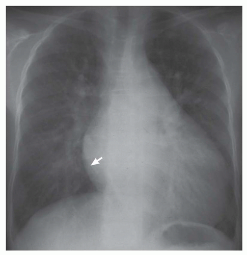
FIG. 31-1. Patent ductus arteriosus. Note pulmonary arterial overcirculation and cardiomegaly. Pulmonary arterial overcirculation is indicated by prominent hilar vessels. There is a left atrial double density (arrow) and enlarged aortic arch.


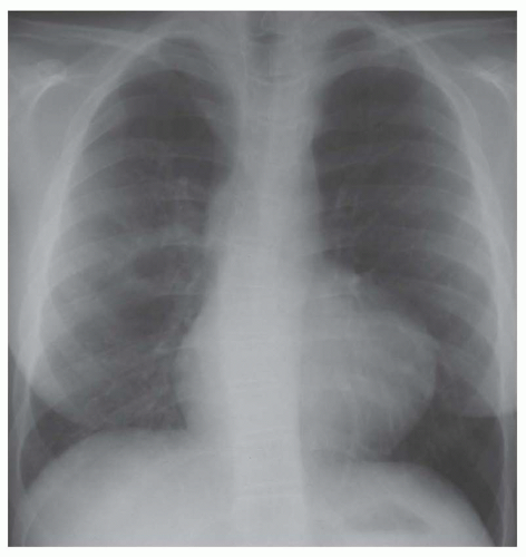
FIG. 31-2. Tetralogy of Fallot. Note decreased pulmonary vascularity without cardiomegaly. The main pulmonary arterial segment is concave and the hilar vessels are small. The apex is situated high above the diaphragm.


Group III: right-to-left shunts with cardiomegaly

Cyanosis

Radiographic evidence of normal or decreased pulmonary blood flow and cardiomegaly (Fig. 31-3).


Group IV: admixture lesions (i.e., both right-to-left and left-to-right shunts)

Cyanosis

Radiographic evidence of increased pulmonary arterial vascularity and usually cardiomegaly (Fig. 31-4).


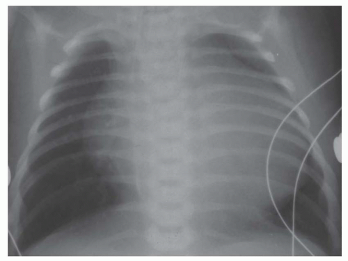
FIG. 31-3. Ebstein’s anomaly. Note decreased pulmonary vascularity with cardiomegaly. Hilar vessels are small and segmental pulmonary arteries are hardly visible, especially in the upper lobes. Vector of enlargement of the apex of the heart is directly lateral, indicating right ventricular enlargement.


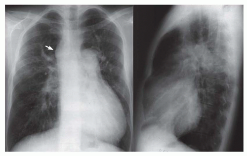
FIG. 31-4. Truncus arteriosus, type I. Note pulmonary arterial overcirculation in the presence of cyanosis and cardiomegaly. There is an enlarged aorta with right arch (arrow).

It is frequently difficult to distinguish between normal and diminished pulmonary vascularity. This observation can be greatly simplified, however, when one remembers that normal pulmonary vascularity, as gleaned from the radiograph in a patient with cyanosis, can be equated with decreased pulmonary vascularity. Consequently, the major observation on the radiograph in terms of pulmonary vascularity in the cyanotic patient is to determine whether the pulmonary vascularity is increased. Normal or diminished pulmonary vascularity in a patient with cyanosis indicates that the lesion produces a right-to-left shunt. Increased pulmonary vascularity in a cyanotic patient indicates that there is an admixture lesion; the cyanosis is indicative of right-to-left shunting, and increased pulmonary vascularity is a sign of left-to-right shunting.


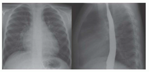
FIG. 31-5. Atrial septal defect. Frontal (left) and lateral (right) views. Pulmonary arterial overcirculation is shown by large hilar and segmental pulmonary arteries. The absence of left atrial enlargement, indicated by no impression on the barium-filled esophagus, is characteristic for an atrial-level shunt.


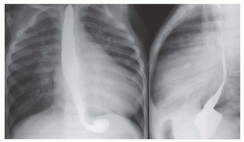
FIG. 31-6. Ventricular septal defect. Frontal (left) and lateral (right) views. Pulmonary arterial overcirculation is evidenced by shunt vessels and prominent hilar vessels. Heart size is increased in proportion to overcirculation. Left atrial enlargement produces impression on and displacement of the barium-filled esophagus, as shown on the lateral view.


GROUPS OF CONGENITAL HEART LESIONS


Group I

Group I contains all of the left-to-right shunts; consequently, this is the group into which most patients with congenital heart disease are classified. The criteria that place a patient within this category are for the most part dependent on the clinical recognition of the absence of cyanosis with the subsequent demonstration on the chest radiograph of increased pulmonary arterial vascularity (see Figs. 31-1 and 31-5,31-6,31-7,31-8 and 31-9). The degree of cardiomegaly is usually in proportion to the increase in pulmonary vascularity. The left-to-right shunts are volume overload lesions. Consequently, there is frequently cardiomegaly, and this cardiomegaly should in general be in proportion to the prominence of the pulmonary vascularity.
When cardiomegaly exists out of proportion to the pulmonary arterial vascularity, then one must consider a number of possibilities. One of the possibilities is that the left-to-right shunt is diminishing in size because of a decrease in the size of the ventricular septal defect (VSD). Another consideration is the coexistence of additional cardiac lesions, such as primary myocardial disease or coarctation of the aorta.


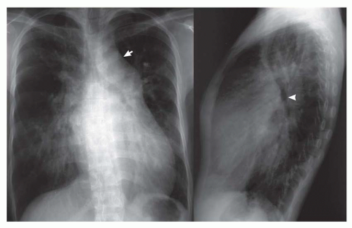
FIG. 31-7. Patent ductus arteriosus. Frontal (left) and lateral (right) views. Note pulmonary arterial overcirculation and cardiomegaly. The prominent aortic arch (arrow) and descending aorta are diagnostic signs of patent ductus arteriosus. On the lateral view, the enlarged left atrium causes posterior displacement of the left bronchus (arrowhead).


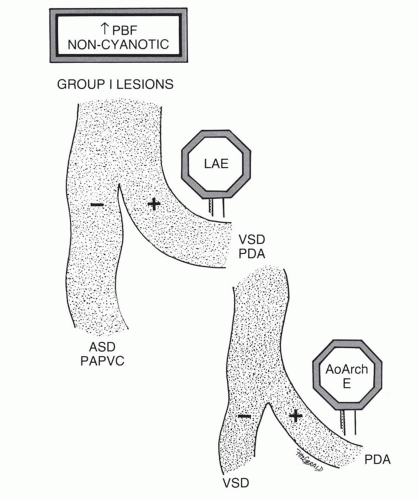
FIG. 31-8. Signposts on the diagnostic pathway of left-to-right shunts.

Two signposts can be used to help distinguish among the various types of left-to-right shunts (see Fig. 31-8). The first of these is the left atrium. Left atrial enlargement indicates that the predominant lesion is not an atrial level shunt but rather a VSD or a patent ductus arteriosus (PDA). The atrial septal defect and partial anomalous pulmonary venous connection lack both of these signposts (see Fig. 31-5). The next signpost is the aortic arch. A prominent aortic arch distinguishes between the PDA and the VSD. The aortic arch usually has a normal dimension or is small in VSD (see Fig. 31-6). PDA is associated with left atrial enlargement and a prominent aortic arch (see Figs. 31-1 and 31-7). In infants, prominence of the aortic arch may be difficult to recognize, so this signpost may not always be available. Consequently, since a VSD is a more frequent lesion, this should be the diagnosis when there is left atrial enlargement and no clearly discernible enlargement of the aortic arch. An exception to this rule is in the premature infant, where a PDA is statistically by far the most frequent congenital heart lesion. The radiograph of the premature infant with PDA usually does not disclose signs of left atrial and aortic arch enlargement.


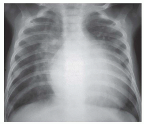
FIG. 31-9. Ventricular septal defects. Large-volume left-to-right shunt causing pulmonary edema, severe pulmonary arterial overcirculation, and cardiomegaly. Indistinct hilar and segmental arteries on the right side are caused by interstitial edema.

The plain radiograph may be useful in determining the severity and progression of left-to-right shunts. The severe volume overload with large left-to-right shunts causes pulmonary venous congestion or pulmonary edema in addition to pulmonary arterial overcirculation (see Fig. 31-9). In individuals with large left-to-right shunts, there should also be substantial cardiomegaly.


Group II

A lesion is included in group II when there is cyanosis and the plain radiograph demonstrates diminished or normal pulmonary vascularity and the absence of substantial cardiomegaly (see Figs. 31-2, 31-10, and 31-11). The pathophysiology that produces this constellation of findings
involves a nonrestrictive intracardiac shunt and a severe obstruction to pulmonary blood flow. The nonrestrictive intracardiac shunt permits equalization of the pressures between two chambers, and this prevents substantial enlargement of the right ventricle. Consequently, there is usually little or no cardiomegaly. An example of the importance of the size of the intracardiac defect is in patients with tricuspid atresia. The patient with tricuspid atresia with a large atrial septal defect demonstrates little or no cardiomegaly (Fig. 31-12). On the other hand, the patient with tricuspid atresia with a restrictive atrial septal defect experiences substantial right atrial enlargement, which results in cardiomegaly. Consequently, the former patient would be classified in group II, the latter patient in group III. Tricuspid atresia can be classified in group IV when there is an associated increase in pulmonary blood flow, which is caused by either a large left-to-right shunt at the ventricular septal level or the concurrence of transposition of the great vessels. Transposition of the great arteries (TGA) occurs in approximately 30% of patients with tricuspid atresia.


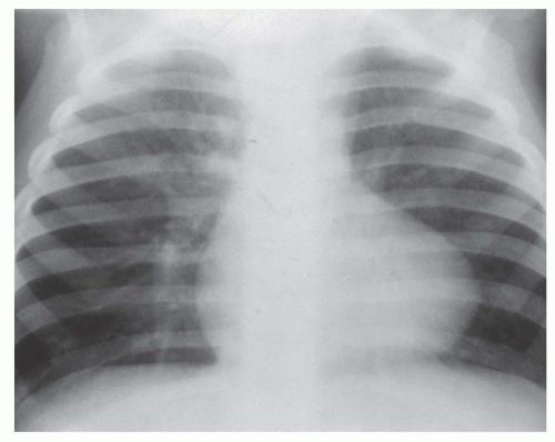
FIG. 31-10. Tetralogy of Fallot. Note pulmonary oligemia with more diminished vascularity on the left, especially the left upper lobe. Normal heart size and concave pulmonary artery segment are characteristic features in the infant.


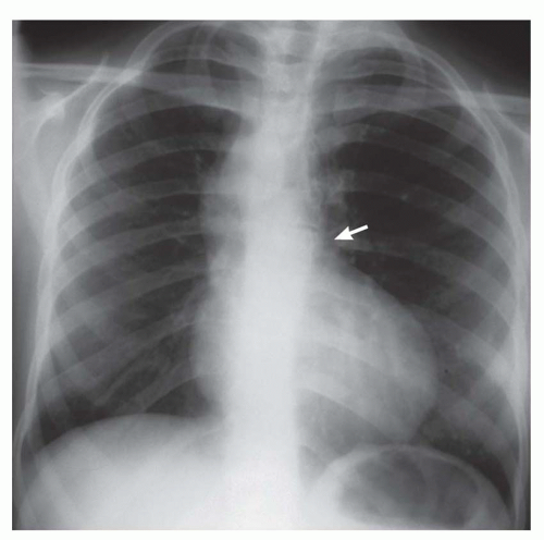
FIG. 31-11. Pulmonary atresia with ventricular septal defect (severe tetralogy of Fallot). Pulmonary oligemia, absent main pulmonary artery segment (arrow), and normal heart size are characteristic features. There is a right aortic arch.


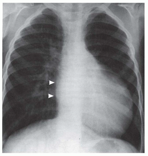
FIG. 31-12. Tricuspid atresia with large (nonrestrictive) atrial septal defect. There is decreased pulmonary vascularity and only mild cardiomegaly. Note the flattened right atrial border (arrows), which is characteristic for this lesion when there is a large nonrestrictive atrial septal defect.

Statistically, the most frequent lesion in group II is tetralogy of Fallot. The remaining diagnostic considerations are, for the most part, variants of tetralogy of Fallot. Some examples of these lesions are TGA with severe pulmonary stenosis and unrestrictive VSD and double-outlet right ventricle with severe pulmonic stenosis and an unrestrictive VSD. Table 31-1 provides a reasonably complete list of the differential diagnostic considerations in group II. However, the plain radiograph infrequently permits a specific diagnosis to be chosen from among this myriad of lesions.


Group III

Group III lesions differ from group II lesions by the radiographic observation of cardiomegaly (see Figs. 31-3 and 31-13,31-4 and 31-15). These patients have cyanosis, normal or decreased pulmonary vascularity, and a substantial degree of cardiomegaly. The cardiac chamber that is frequently enlarged in this lesion is the right atrium. Many of the patients in this category have substantial tricuspid regurgitation, which is a major pathogenetic mechanism of the right atrial enlargement and cardiomegaly. The “wall-to-wall” heart (extension from the right to the left chest wall) should
prompt the diagnostic consideration of a lesion causing tricuspid regurgitation.


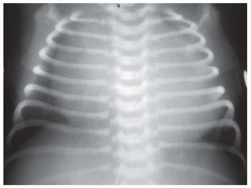
FIG. 31-13. Pulmonary atresia with intact ventricular septum, type II. Substantial tricuspid regurgitation in association with this anomaly (type II) causes right-sided chamber enlargement, especially right atrial enlargement.


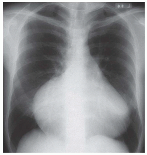
FIG. 31-14. Ebstein’s anomaly in an adult. Note decreased pulmonary vascularity and marked cardiomegaly. The prominent bulging and elongation of the right heart border are indicative of severe right atrial enlargement.

There is no statistically dominant diagnostic consideration in this category, but the following lesions must be considered in the differential diagnosis: severe (“critical”) pulmonary stenosis with an atrial septal defect or patent foramen ovale; type II pulmonary atresia with intact ventricular septum; tricuspid atresia with a restrictive atrial septal defect; and Ebstein’s anomaly. In the older child and adult with this constellation of findings, the most likely diagnosis is Ebstein’s anomaly (see Fig. 31-14). Uhl’s anomaly is a rare cause of a cardiac configuration similar to Ebstein’s anomaly. Another unusual diagnosis in this category, which appears only in the neonatal period, is tricuspid regurgitation of the newborn (see Fig. 31-15). In this entity there is frequently substantial cardiomegaly, diminished pulmonary blood flow, and cyanosis within the first few days of life. However, with reduction in pulmonary vascular resistance over time, the amount of tricuspid regurgitation decreases and the cardiomegaly may resolve.


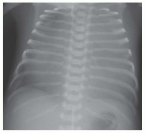
FIG. 31-15. Tricuspid regurgitation of the newborn. Pulmonary vascularity is decreased and marked cardiomegaly is present due to right-sided chamber enlargement. Extreme cardiomegaly producing the “wall-to-wall” heart is usually due to severe tricuspid regurgitation.


Group IV

A lesion is included in this group when the radiograph displays pulmonary arterial overcirculation in the presence of cyanosis. The heart size is usually increased. The observation of increased pulmonary vascularity in a patient with cyanosis is an incongruous finding and should alert the observer to the presence of an admixture lesion rather than a strictly leftto-right shunt. An aid to remembering the major diagnoses in this category is the letter T. The most common diagnosis in this category is TGA, which is the most frequent cyanotic congenital heart lesion at birth (Fig. 31-16). The other diagnostic considerations are truncus arteriosus (Fig. 31-17),
total anomalous pulmonary venous connection (Fig. 31-18), tricuspid atresia, and single (“tingle”) ventricle. Doubleoutlet right ventricle and double-outlet left ventricle are also considered in this category, but these can be brought to mind when one thinks of TGA, since these lesions are essentially hybrids of TGA. The lesion that is frequently forgotten in this group is multiple pulmonary arterial venous malformations. The patient with multiple pulmonary arterial venous malformations is frequently mildly or even moderately cyanotic, and because of the several malformations within the lung, there is the appearance of increased pulmonary arterial vascularity.


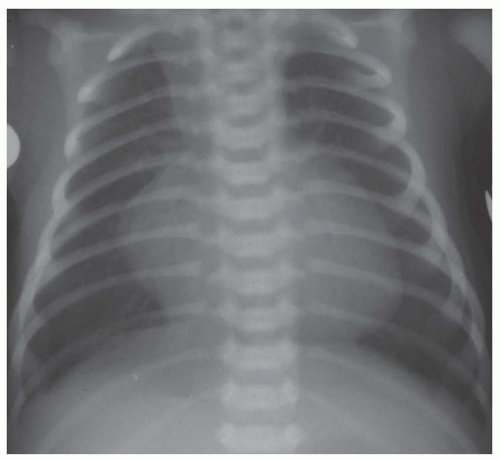
FIG. 31-16. Transposition of the great arteries. Pulmonary arterial overcirculation and an ovoid heart with a narrow base (vascular pedicle) of the heart are characteristic features.


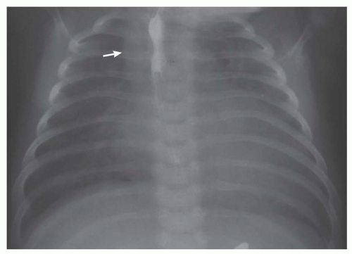
FIG. 31-17. Truncus arteriosus. Pulmonary arterial overcirculation and right aortic arch (arrow) are characteristics of truncus arteriosus.


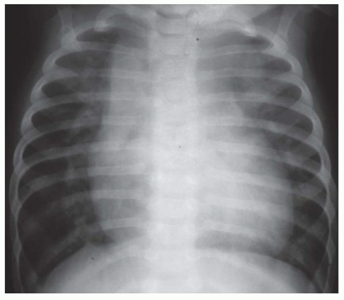
FIG. 31-18. Total anomalous pulmonary venous connection, supracardiac type (type I). Note pulmonary arterial overcirculation and cardiomegaly. Enlargement of supracardiac region is caused by an enlarged left-sided vertical vein and a dilated right superior vena cava; it is characteristic of this anomaly.


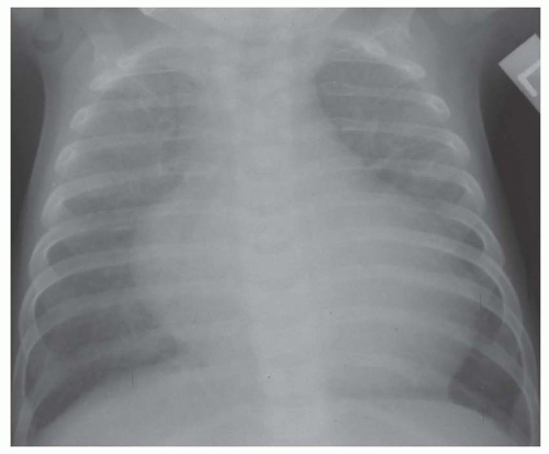
FIG. 31-19. Endocardial fibroelastosis. Pulmonary edema and cardiomegaly are characteristic features of group V lesions.


PULMONARY VENOUS CONGESTION OR PULMONARY EDEMA

A fifth group of congenital lesions are those that produce predominantly pulmonary venous congestion and alter the pulmonary venous vascularity rather than the pulmonary arterial vascularity. Patients with these lesions may have shunts, but inclusion in group V requires that the predominant pathophysiologic event is pulmonary venous congestion (Figs. 31-19,3-20 and 31-21; see Table 31-2).

The clinical features of the group V lesions are lack of cyanosis and frequently severe symptoms of heart failure. These
usually consist of dyspnea, tachypnea, and tachycardia. The salient radiographic findings are indistinctness of the pulmonary vascularity, especially in the perihilar area, or interstitial pulmonary edema (see Figs. 31-19 and 31-21). Another observation that places a lesion into this group is disproportionately prominent cardiomegaly in comparison to the prominence of pulmonary vascularity (see Figs. 31-19 and 31-20).


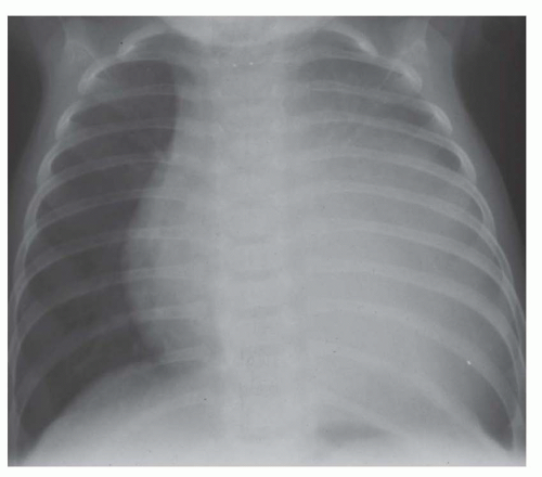
FIG. 31-20. Anomalous origin of the left coronary artery from the pulmonary artery. Cardiomegaly is disproportionate to pulmonary vascularity in a noncyanotic infant. Left atrial enlargement (right retrocardiac double density) is caused by mitral regurgitation from papillary muscle infarction.


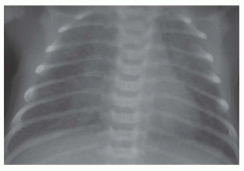
FIG. 31-21. Total anomalous pulmonary venous connection, type III. Radiograph shows pulmonary edema and normal heart size.

The lesions included in this category are listed in Table 31-2. The statistical frequencies of the lesions in this category are also important in deciding on the diagnosis. Diagnosis in this category includes conditions that produce reversible stresses upon the heart of the newborn as well as structural cardiac lesions. Lesions in this category tend to present at certain times after birth; for instance, the nonstructural causes of pulmonary venous congestion or edema usually present within the first day or two of life. Abnormalities that may be encountered within the first day of life include severe anemia (hydrops fetalis), asphyxia, hypocalcemia, hypoglycemia, abnormalities of heart rate and rhythm, hypervolemia, and intrauterine myocarditis. Pulmonary venous congestion with substantial cardiomegaly presenting in the first day or so of life is a feature of hypoplastic left heart (Fig. 31-22). Pulmonary venous congestion with an essentially normal heart size presenting within the first day or so of life is the feature of total anomalous pulmonary venous connection, infradiaphragmatic type, with obstruction (see Fig. 31-21). In the infant presenting with these features between 1 and 3 weeks of age, statistically the most frequent diagnosis is coarctation of the aorta (Fig. 31-23). Rib notching is not evident in infants with coarctation.


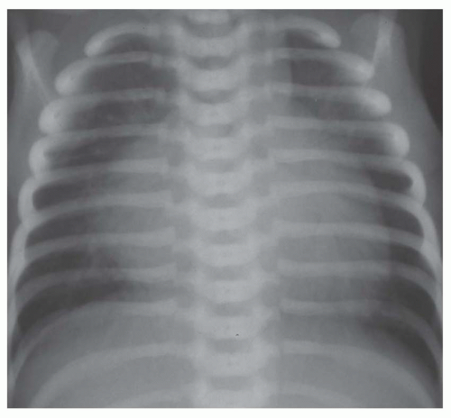
FIG. 31-22. Hypoplastic left heart. Note pulmonary venous congestion and edema and cardiomegaly. Prominent right atrium and ventricle and posterior aortic arch are characteristic features of this lesion.


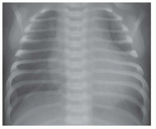
FIG. 31-23. Severe coarctation of the aorta in a newborn. There is marked cardiomegaly with pulmonary edema.


SALIENT RADIOGRAPHIC FEATURES OF SPECIFIC LESIONS


Acyanotic


Atrial Septal Defect and Partial Anomalous Pulmonary Venous Connection

There are four types of atrial septal defects: secundum (most frequent); primum; sinus venosus (superior and inferior vena caval locations); and coronary sinus (least frequent). The primum type is usually part of an atrioventricular septal defect (AVSD), which was formerly called endocardial cushion defect. In addition, a patent foramen ovale exists in many children with congenital heart disease, and the foramen may be stretched in the setting of elevated right-sided pressures. An aneurysm may also form at the site of the thin fossa ovalis; this may occur as an isolated anomaly or may exist in association with a septal defect or patent foramen ovale. The defects are named according to their position in the atrial septum: ostium secundum in the region of the fossa ovalis, which is approximately the middle of the septum; primum in the lower part of the septum and bordering on the atrioventricular valves; sinus venosus in either the upper part of the septum and bordering on the ostium of the superior
vena cava or in the lower septum and bordering on the ostium of the inferior vena cava. A rare type of defect occurs at a site normally occupied by the coronary site and coexists with absence of the wall separating the coronary sinus from the left atrium so that the associated left superior vena cava enters into the left atrium. The coexistence of large primum and secundum defects constitutes a common atrium.


TABLE 31.3 Salient Radiographic Features of Atrial Septal Defect


Pulmonary arterial overcirculation: Generally a 2:1 shunt must exist before pulmonary plethora is universally present. About 50%-60% of patients with less than 2:1 shunt have only mild or no evident pulmonary plethora. Pulmonary edema rarely occurs in the simple atrial septal defect.


Enlargement of right atrium (see Fig. 31-5)


Enlargement of right ventricle


Enlargement of main and hilar pulmonary arterial segments: In older subjects, the right pulmonary artery is sometimes especially prominent (see Fig. 31-24).


Small ascending aorta and aortic arch


Small superior vena caval shadow


The isolated atrial septal defect and partial anomalous pulmonary venous connection conduct left-to-right shunts. A stretched foramen ovale or atrial septal defect may permit predominant right-to-left shunting in complex lesions with severe right-sided obstruction (i.e., tricuspid atresia). The volume of shunting across an interatrial communication usually depends on the size of the defect and the relative distensibility of the two ventricles. The wall of the right ventricle is more distensible than the left ventricle during diastole, so blood preferentially flows toward the right ventricle at this time. However, obstruction of flow into or out of the right ventricle can reverse this pattern. A large atrial septal defect is defined as one that results in equalization of pressure between the atria (Tables 31-3,31-4 and 31-6; Figs. 31-24 and 31-25).


Atrioventricular Septal Defect (Endocardial Cushion Defect)

The embryonic endocardial cushions contribute to the development of the medial portions of the mitral and tricuspid valves, the primum atrial septum, and the inlet portion of the ventricular septum (Fig. 31-26). Defects in this region have been called endocardial cushion defects but more recently have received the name atrioventricular septal defects (AVSDs). The fundamental lesion is a common atrioventricular valve and variable deficiency of the primum atrial septum and the inlet ventricular septum. The atrioventricular valve in this anomaly has five leaflets, with two of the leaflets spanning the ventricular septum and the opening to both ventricles. The spanning leaflets are the anterior and posterior bridging leaflets. If there is a tongue of tissue connecting the anterior and posterior bridging leaflets and this tongue is attached to the crest of the inlet ventricular septum, then incomplete forms of the defect result. The anomaly exists in a complete form with a single atrioventricular valve, primum atrial septal defect, and inlet VSD. In the complete form, no connecting tissue exists between the bridging leaflets. Incomplete forms are said to exist when there are two atrioventricular valves; the individual valves are formed by the connecting tongue of tissue. Portions of the valves are frequently deficient, such as underdevelopment of the septal leaflet of the tricuspid valve and a cleft in the anterior leaflet of the mitral valve. Actually, this “cleft” is the commissure between the anterior bridging leaflet and the mural leaflet of the left-sided portion of the atrioventricular valve.


TABLE 31.4 Salient Radiographic Features of Atrial Septal Defect with Pulmonary Arterial Hypertension


Enlarged main and central pulmonary arteries (see Fig. 31-24).


Disparity in enlargement of central and lobar arteries to peripheral arteries.


Calcification of main or central pulmonary arteries.


Onset of severe pulmonary arterial hypertension may be associated with reduction in degree of cardiomegaly or normal heart size.


Cardiomegaly may be persistent because of tricuspid insufficiency caused by severe pulmonary hypertension.


TABLE 31.5 Salient Radiographic Features of Partial Anomalous Pulmonary Venous Coarctation


Pulmonary arterial overcirculation: This may be apparent or more severe only in the lung with anomalous drainage.


Enlargement of right atrium


Enlargement of right ventricle


Enlargement of main and hilar pulmonary arterial segments


Small ascending aorta and aortic arch


Enlargement of superior vena cava, azygous vein, coronary sinus or other systemic veins, depending on site of connection


Prominent left superior vena cava


Abnormal course of pulmonary veins through the lung or in relation to mediastinal margins (see Fig. 31-25)


The most common incomplete lesion is a primum atrial septal defect and a “cleft” in the mitral valve, causing varying degrees of mitral regurgitation. Because the primum atrial septal defect is situated immediately above the cleft, mitral regurgitation may traverse the defect and enter the right atrium. Consequently, the left atrium may not be enlarged
even in patients with substantial mitral regurgitation (Table 31-7; Figs. 31-27 and 31-28).


TABLE 31.6 Salient Radiographic Features of Scimitar Syndrome


Enlarged curved vascular structure coursing medially toward the right diaphragm: This structure enlarges in diameter as it approaches the diaphragm (see Fig. 31-25).


Dextroposition of the heart


Hypoplasia of right lung


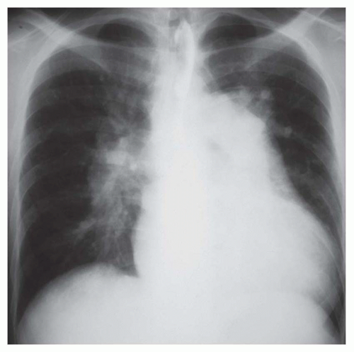
FIG. 31-24. Atrial septal defect in an adult. The radiograph shows pulmonary arterial overcirculation and cardiomegaly due to right-sided chamber enlargement. Severe dilatation of the central pulmonary arteries is a feature of this anomaly in the adult. The right pulmonary artery is very prominent.


Ventricular Septal Defect

VSDs have been characterized by their location in the septum: perimembranous, outlet, inlet, and trabecular (Fig. 31-29). Defects in the perimembranous and outlet regions have also been described in relation to the crista supraventricularis of the right ventricle as infracristal (more frequent) and supracristal types. While any perimembranous or outlet VSD can cause aortic regurgitation, the supracristal type frequently causes prolapse of the right sinus of Valsalva and aortic regurgitation. Prolapsed sinus tissue may reduce the size or obliterate the septal defect. The outlet defect may be caused
by malposition of the outlet septum, resulting in a small right ventricular outflow region and an aorta overriding the septal defect (tetralogy of Fallot). VSDs are not uncommonly multiple. Multiple defects in the trabecular septum may produce a “Swiss cheese septum.”


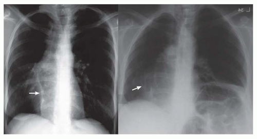
FIG. 31-25. Two patients with scimitar syndrome. Left: Radiograph showing a scimitar vein near the right diaphragm, a dextroposed heart, and a small right lung. The scimitar vein (arrow) enlarges in its course toward the diaphragm. Right: Radiograph showing multiple anomalous veins (arrow) arching toward the right hemidiaphragm. The increased diameter of the veins from superior to inferior indicates that they are anomalous veins rather than pulmonary arteries. The heart is dextroposed. There is an incidental eventration of the left hemidiaphragm.


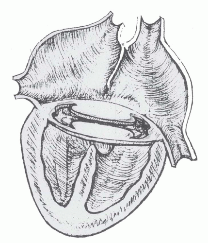
FIG. 31-26. Diagram of complete form of atrioventricular septal defect. The defect consists of a primum atrial septal defect, an inlet ventricular septal defect, and a single atrioventricular valve spanning the ventricular septal defect.


TABLE 31.7 Salient Radiographic Features of Atrioventricular Septal Defect


Skeletal features of trisomy 21, such as 11 ribs, double manubrial ossification center, and tall vertebral bodies


Pulmonary arterial overcirculation: This is severe in the complete forms and may be associated with pulmonary edema. Concurrent pneumonia is frequent in the complete form, especially in the child with mongolism.


Enlargement of right atrium: The superior margin of the right atrium is frequently prominent (see Figs. 31-27 and 31-28).


Enlargement of right ventricle (see Fig. 31-28)


Enlargement of main and central pulmonary arterial segments


Left atrial enlargement may be present but is generally not severe and may be absent despite mitral regurgitation.


Small thoracic aorta


A cleft mitral valve without a primum defect (rare) produces the radiographic configuration of mitral regurgitation.

Only gold members can continue reading. Log In or Register to continue

Stay updated, free articles. Join our Telegram channel

Oct 10, 2016 | Posted by in CARDIOVASCULAR IMAGING | Comments Off on Radiography of Congenital Heart Disease

Full access? Get Clinical Tree

Get Clinical Tree app for offline access