and Zdeněk Fryšák1
(1)
Department of Internal Medicine III – Nephrology, Rheumatology and Endocrinology, Faculty of Medicine and Dentistry, Palacky University Olomouc and University Hospital Olomouc, Olomouc, Czech Republic
Keywords
Thyroid hemiagenesisLipomaBranchial cleft remnantsDermoid cystCellulitis—phlegmonPostoperative hemorrhageThrombus in the internal jugular vein21.1 Essential Facts
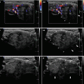
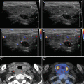
Thyroid hemiagenesis (Fig. 21.1aa) is a rare congenital abnormality of the thyroid gland, characterized by the absence of one lobe. The true prevalence of this congenital abnormality is not known because the absence of one thyroid lobe usually does not cause clinical symptoms by itself. In 2000, Shabana et al. performed a systematic US study of the thyroid gland volume for the evaluation of iodine deficiency in 2845 normal Belgian schoolchildren and found an absence of the left lobe in six children (four girls and two boys). There was no association with other thyroid malformations or dysfunction. The prevalence of thyroid hemiagenesis was established of 0.2% and confirmed the female predominance and higher incidence of agenesis of the left lobe [1].
A large retrospective study between 1970 and 2010 in Sicily by De Sanctus et al. found 329 (1.3%) cases of thyroid hemiagenesis in 24,032 unselected schoolchildren from 11 to 14 years old. It is interesting to note that most cases have an agenesis of the left lobe (80%) followed by the isthmus (44–50%), and the girl-to-boy ratio was 1:1.4, contrary to other studies that document a higher prevalence in women. The hemiagenesis is mostly diagnosed when a patient presents a lesion in the remaining/functioning lobe. The solitary functioning lobe can be a site of pathological changes similar to a normally developed thyroid gland [2].
Lipomas arise in almost 50% of all soft tumors. The neck lipomas are rare tumors that may present as painless masses with slow growth in the lateral portions of the neck [3].
Branchial cleft remnants, which arise from the incomplete obliteration of any branchial tract, accounts for the majority of branchial cleft anomalies. Clinically they can present as different morphologic patterns: approximately 70% are hypoglossal duct sinuses and cysts, 25% are branchial cysts and sinuses, and 5% are cystic hygromas [4].
Branchial cleft cysts (BCCs) can first come to clinical attention usually in a child or young adult in the second through fourth decades of life. They present clinically as smooth, soft, ovoid, cystic masses in the lateral part of the neck, behind the angle of the mandible, and anterior to the sternocleidomastoid muscle [5, 6].
Dermoid cysts (DCs) are benign lesions histologically composed of tissues originating from the ectoderm and the mesoderm, but never from the endoderm. Approximately 7% of all DCs are seen in the head and neck area (the third most common site, after the coccyx in 44% and the ovary in 42%), and they are located most often in superficial subcutaneous tissues. DCs can occur at any age with no gender predilection, but more than 50% of all cases are detected by the age of 6 years. DCs are generally asymptomatic unless they grow enough to be conspicuous, causing cosmetic problems or compressive effects. Congenital cysts arise from a rest of embryonic epithelium such as bronchial cleft cyst, and are acquired as a result of traumatically implanted skin in the deeper tissues. The lumen of the cyst contains amorphous material with a few hair shafts [7].
Other non-thyroid cystic neck masses include lymphatic malformation, ranula, and parathyroid cyst (Fig. 22.10aa). US-guided sclerotherapy (by sclerosant agents 96% ethanol or OK-432) is an alternative treatment for all benign non-thyroid cystic neck masses [8].
Intravascular extension of tumor cells by direct intraluminal spread is more commonly encountered in anaplastic thyroid carcinoma. Invasion of internal jugular vein (IJV) by thyroid cancer indicates a poor outcome. Differentiated thyroid carcinomas (DTC) as papillary and more often follicular and Hurthle cell carcinomas have well-documented microscopic characteristics of microinvasion affecting the great cervical veins. Direct extraluminal vascular invasion with DTCs is extremely rare. Cervical and arm edema and pain are the most common presentations of IJV thrombosis [9, 10].
The cellulitis—phlegmon and superficial abscess are acute processes of neck swelling. Clinically, there are general differences between the two. The pain caused by cellulitis tends to be more severe and generalized than the localized pain associated with an abscess. Cellulitis often presents with swelling, warmth, erythema, and tenderness over the affected area and equates to the descriptive Latin terms “tumor,” “calor,” “rubor,” and/or “dolor.” The firmness of the cellulitis can range from doughy to indurate. The borders of cellulitis are typically large, smooth, ill-defined, and do not contain pus. An abscess usually has a small and well-circumscribed borders with signs of inflammation, and is soft or fluctuant to palpation, indicating a pus-filled cavity. Patients with systemic infections often have fever [11].
Differentiating a soft tissue abscess from cellulitis is important because each of these disorders requires a different treatment approach. Whereas cellulitis responds to systemic antibiotics, an abscess must be drained surgically. Diagnosis is complicated by the fact that abscesses often begin as cellulitis and therefore the two conditions frequently coexist [12].
Squire et al. investigated 135 patients with soft tissue infection (76 with abscess and 59 with cellulitis). Comparison of clinical examination alone with US examination for superficial abscess showed the sensitivity 86% vs. 98% and the specificity 70% vs. 88%, the positive predictive value 81% vs. 93%, and the negative predictive value 77% vs. 97%. In 18 cases, US was not consistent with the clinical examination, but US finding was correct in 17 (94%) of the cases. However, three patients with false-positive findings of abscess on US turned out to have hematomas. Concluding, bedside US improves accuracy in detection of superficial abscesses [12].
Postoperative hemorrhage is a well-known complication of thyroid surgery. It requires special attention because it may represent a life-threatening situation due to acute airway obstruction from the deep hematoma. The reported incidence of post-thyroidectomy hemorrhage varies from 0.36 to 4.3%. The etiology of post-thyroidectomy hemorrhage has been described as slipping of the major vessel ligature, reopening of the cauterized veins, retching and bucking of the patient during recovery, a Valsalva maneuver, increased blood pressure and oozing from the thyroid cut area. Neck swelling is frequently identified as a representative clinical sign of post-thyroidectomy hemorrhage. The ecchymosis is often identified in cases of superficial hematoma, but rare in cases of deep hematoma [13].

Fig. 21.1
(aa) A 71-year-old woman with hemiagenesis of the RL and isthmus, Hashimoto’s thyroiditis (HT) in the solitary LL. The diagnosis was established in middle-age, when she presented with features of hypothyroidism and anti-thyroid antibodies positive for HT. US appearance: empty right thyroid bed; LL—inhomogeneous with hypoechoic micronodular structure and thin hyperechoic fibrous septa; volume 5 mL; transverse
21.2 US Findings of Lesions at the Anterior Neck Space Mimicking Thyroid
US findings of dermoid and epidermoid cysts (Fig. 21.6aa): cyst has an internal echo, with a solid appearance with only slight or no posterior echo enhancement. Amorphous keratinous debris from keratinizing stratified squamous epithelium fills the lumen of each cyst, producing the internal echoes. Caution! Lipomas are indistinguishable from these cysts. US-FNAB clarifies the cystic nature, cytological evaluation reveals benign-appearing squamous cells and amorphous cellular debris [14].
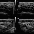
US findings of lipoma (Fig. 21.3bb): an elongated isoechoic or hyperechoic mass in the subcutaneous tissues (very much like those of subcutaneous fat tissue). The existence of striated echoes in the tumor corresponding to the septa increases the possibility of lipoma. US is useful for acquiring information about the nature, size, and depth of the lesion as well as its relationship to adjacent vessels and other structures, especially the thyroid gland [3]. Caution! US can reveal lipoma and coexisting MNG (Fig. 21.4aa).
US findings of a thrombus in the internal jugular vein (Fig. 21.7aa): the vein is expanded and contained a thrombus of heterogeneous, dominantly hyperechoic structure [15].
US findings of a tumor thrombus in the internal jugular vein (Fig. 15.36aa): cancer mass of thyroid lobe (often with multiple areas of necrosis) extending and invading the jugular vein wall; the cancer and thrombus are hypervascular in CFDS. Moreover, CFDS demonstrates a totally or partially absent flow in IJV [9].
US findings of superficial abscess: spherical in shape with poorly defined borders, content forms heterogenic, anechoic, or hypoechoic mass containing a variable amount of internal echoes. Compression of the mass with the transducer may demonstrate movement or “swirling” of pus [12].
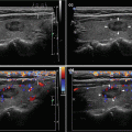
US findings of cellulitis—phlegmon (Fig. 21.9aa): a thickening and diffuse hyperechogenicity of the subcutaneous fat with obliteration of the interface between the echogenic fat and the dermis. This is commonly referred to as “cobblestoning.” There are increased local reactive LNs. CFDS helps differentiate soft tissue infections by demonstrating diffuse hypervascularity in areas of inflammation [12].
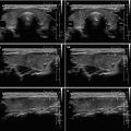
US findings of hematomas (Fig. 21.8dd): the echogenicity of blood contained within a hematoma is variable and depends largely on time of stasis. Initially blood appears anechoic, but over several hours, as the blood begins to coagulate, it appears more and more echogenic. Pus also appears echogenic but is differentiated from congealed blood by its heterogeneity. Clotted blood appears uniformly echogenic, while pus has a variable echogenic pattern [12]. Superficial hematoma indicates a hematoma located between the subplatysmal dissection plane and the strap muscles, while deep hematoma indicates a hematoma between the strap muscles and the thyroid bed [13].
21.3 US Findings of Artifacts [17]
Edge shadowing from thyroid cystic lesion at tangential angle—“ears” (similarly hepatic cysts, gall-bladder); see more in Chap. 7 (Fig. 7.10cc).
So-called “comet tails” as bright hyperechoic reverberation artifacts (the condensed colloid proteins); see more in Chap. 7 (Fig. 7.9aa).
Posterior acoustic enhancement behind cyst (similarly hepatic cysts, gall-bladder); see more in Chap. 7 (Fig. 7.4aa).
A “mirror-image” artifact arises when there is a highly reflective surface. Typically hepatic cyst is reflected back to the highly reflective surface, e.g. diaphragm. A similar situation may occur in locating a cystic nodule in the midline in the isthmus (Fig. 21.12aa), and a “mirror-image” can be seen beyond the tracheal wall (the most hyperechoic arc-shape line is the interface of the tracheal wall and air in the tracheal lumen).
Stay updated, free articles. Join our Telegram channel

Full access? Get Clinical Tree


