Readout and Background Suppression Techniques
David Thomas
Matthias Günther
Xavier Golay
Arterial spin labeling (ASL) is a low-sensitivity method. As explained in Chapter 15, the general signal expected in ASL is only a fraction of the original magnetic resonance imaging (MRI) signal. As such, numerous schemes have been proposed to optimize the signal-to-noise ratio (SNR) in ASL techniques. For this reason, two main areas of developments have been pursued in the past 2 decades. on the one hand, many efforts were invested in the optimization of rapid and high SNR readouts, allowing the acquisition of a large number of averages over a few minutes to ensure at least a minimum SNR of 1500:1 to 2000:1, which in turn leads to a typical SNR of 10:1 in perfusion maps. Most methods used in ASL so far have been based on rapid gradient-echo imaging schemes, such as echo-planar imaging (EPI) or spiral imaging, while recent developments have also seen the pursuit of other high SNR methods, in particular in combination with three-dimensional acquisition schemes.
The second line of research has been the development of techniques to reduce unwanted signal from the tissue of interest to an absolute minimum by using a series of inversion pulses to selectively minimize the static tissue signal. These methods are known as background suppression (BS) techniques. BS is of particular importance with multishot acquisitions because even small motion during data acquisition could deteriorate the already very small detectable ASL signal.
A third factor that affects SNR is the generation of the label (continuous, pulsed, pseudo-continuous) and its efficiency in itself. This chapter will summarize the developments made in the past 2 decades for both background suppression and image formation.
Readout Methods for Arterial Spin Labeling
Because of the independence of the spin labeling process and image acquisition, it is possible to combine any of the myriad ASL labeling modules with any MRI acquisition technique. In the very first implementation of ASL, a simple two-dimensional spin-echo acquisition was used to demonstrate the principle.1 But it soon became clear that to achieve an acceptable SNR within a reasonable imaging time, fast imaging techniques would be essential for ASL to succeed. For the first application in humans, single-shot EPI was the logical choice because of its single-shot nature and high SNR efficiency. Nevertheless, even from the early days of ASL, the relative pros and cons of different fast imaging methods have meant that for particular applications, other acquisition schemes might offer significant advantages over EPI. The imaging techniques that have been implemented with ASL, and the specific strengths and weaknesses they may offer, are summarized in this chapter.
Echo-Planar Imaging
Currently, EPI is offered as a standard product sequence by all major MRI manufacturers and is the workhorse sequence for blood oxygenation–level dependent (BOLD) functional MRI (fMRI) and dynamic susceptibility contrast perfusion imaging. As such, a significant amount of effort has been invested in optimizing image quality and minimizing the characteristic artifacts associated with the sequence. ASL can take advantage of this by using EPI as its imaging module. Even before EPI was a well-developed product sequence, it was used in many of the first implementations of ASL for imaging the human brain.2,3,4,5 EPI provides a two-dimensional multislice acquisition scheme that can achieve whole brain coverage in a reasonable time (typically about 50 milliseconds per slice, depending on the image resolution), and this can be reduced with the use of parallel imaging methods, although with the associated SNR penalty.6 EPI has a high SNR efficiency, because it acquires the entire two-dimensional k-space after a single, high flip angle radiofrequency (RF) excitation pulse (usually 90°).
The issues of EPI have been well documented.7 Modern scanners have correction schemes that almost entirely eliminate the appearance of so-called Nyquist or FOV (field of view)/2 ghosts,8 which result from uncorrected gradient-induced phase errors between the odd and even echoes of the EPI readout train. The problem of image distortion in regions of main field (B0) inhomogeneity remains, although various approaches to correct these distortions have emerged in recent years.9 Gradient-echo EPI (GE-EPI) is most widely used with ASL because it minimizes the acquisition time, allowing rapid multislice coverage. In order to keep the BOLD contrast to a minimum (which would otherwise introduce a confounding source of signal intensity change), the shortest possible echo time (TE) is typically chosen, to avoid significant T2* weighting of the sequence. Even when making an attempt to keep TE short with EPI, it can still be significant
(>10 milliseconds) because of the nature of the EPI readout trajectory, which can affect the measured signal in situations when T2* relaxation times are short, such as seen at higher magnetic field strengths (e.g., 7 T). In addition, the use of crusher gradients to eliminate vascular signal might further prolong the minimum TE to at least 20 milliseconds, thereby increasing the susceptibility of EPI to confounding BOLD effects. To reduce BOLD sensitivity for EPI, parallel imaging methods can be applied, which enables shorter TE to be used. Alternatively, spin-echo EPI has also been used as a way to reduce BOLD weighting in ASL; this is particularly suitable for applications to fMRI10 and in preclinical studies where rapid multislice coverage is not essential.11
(>10 milliseconds) because of the nature of the EPI readout trajectory, which can affect the measured signal in situations when T2* relaxation times are short, such as seen at higher magnetic field strengths (e.g., 7 T). In addition, the use of crusher gradients to eliminate vascular signal might further prolong the minimum TE to at least 20 milliseconds, thereby increasing the susceptibility of EPI to confounding BOLD effects. To reduce BOLD sensitivity for EPI, parallel imaging methods can be applied, which enables shorter TE to be used. Alternatively, spin-echo EPI has also been used as a way to reduce BOLD weighting in ASL; this is particularly suitable for applications to fMRI10 and in preclinical studies where rapid multislice coverage is not essential.11
 FIGURE 16.1. Typical brain images when acquired with a pulsed sequence using an echo-planar imaging readout. Arterial spin labeling module quantitative imaging of perfusion using a single subtraction, second version, with TI1 = 800 milliseconds and TI2 = 1800 milliseconds; image resolution 3.9 × 3.9 × 3.5 mm3. TI, inversion time. |
An example of perfusion-weighted ASL brain images using an EPI readout approach is shown in Figure 16.1.
Two-Dimensional Gradient-Echo and TurboFLASH
One of the early implementations of ASL in humans used an acquisition in which short continuous ASL (CASL) pulses were interleaved within the repetition time (TR) of a refocused gradient-echo acquisition,12 although this was soon superseded by snapshot EPI techniques. In preclinical studies, TurboFLASH has been used as the imaging module for ASL to avoid the aforementioned issues associated with EPI at higher field strengths.13,14,15 A combination of short TR (<10 milliseconds) and low flip angle (<20°) is required to keep the image acquisition reasonably short, at the expense of reducing the overall SNR. Note that to maintain the correct ASL contrast, center-out phase encoding should be used. With the advent of human 7T scanners, there has been some reinvestigation of the potential of TurboFLASH ASL,16 although the inherent reduction in SNR that is associated with the low flip angle excitation is likely to be a limiting factor for most ASL applications.
An example of ASL perfusion images of a rodent brain, which was acquired using a TurboFLASH readout, is shown in Figure 16.2.
 FIGURE 16.2. Example of arterial spin labeling (ASL) images acquired using a TurboFLASH readout. Pseudo-continuous ASL perfusion images at 7T (top) and 3T (bottom) at three different resolutions. (Reproduced with permission from Zuo Z, Wang R, Zhuo Y, et al.10 Turbo-FLASH based arterial spin labeled perfusion MRI at 7 T. PLoS One. 2013;8(6):e66612.) |
Look-Locker
Look-Locker (LL) acquisition schemes consist of multiple acquisitions performed following a single spin preparation (inversion).17,18,19 In the context of ASL, this corresponds to the acquisition of several images after a single labeling pulse. The advantage of this type of approach is that it has excellent time efficiency for acquiring ASL images with a
range of post-labeling delay times. It is compatible with both TurboFLASH and EPI imaging modules and has been used for a variety of applications. However, the LL readout also affects the label (i.e., a steady state is reached if a constant readout is applied following a single, upfront label). In other words, the label-control signal at time T will be different if LL readouts are used versus just waiting until time T has elapsed to read out the labeled signal.
range of post-labeling delay times. It is compatible with both TurboFLASH and EPI imaging modules and has been used for a variety of applications. However, the LL readout also affects the label (i.e., a steady state is reached if a constant readout is applied following a single, upfront label). In other words, the label-control signal at time T will be different if LL readouts are used versus just waiting until time T has elapsed to read out the labeled signal.
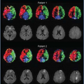
 FIGURE 16.3. (A) T2-weighted anatomical images that demonstrate advanced white matter microvascular lesions in a 92-year-old patient suffering from bilateral narrowing of both common carotid arteries. (B) Calculated cerebral blood flow (CBF) images. (C) Calculated arterial blood volume images. (D) Calculated bolus arrival time to the capillaries (brighter means later arrival times). In spite of the extensive white matter lesions, this patient seems to have maintained a good blood supply and does not present with large neurovascular deficit, apart from the posterior right parietal area, showing a slight decrease in CBF and compensatory increase in arterial blood volume. |
An early implementation of ASL proposed the use of an LL TurboFLASH acquisition after slice-selective and nonselective inversion pulses.20 This approach has since been used quite extensively to measure myocardial perfusion in the rodent heart.21,22,23,24,25 Perfusion quantification is usually based on a simple T1-recovery model; recently, however, the deficiencies of this approach have been realized and a more appropriate model has been proposed that uses a simultaneously measured arterial input function to improve the accuracy of the perfusion values.26
For application to the human brain, LL ASL acquisitions have focused on using EPI as the imaging method. Following the initial proposal of this technique,17 some groups developed and refined the acquisition and postprocessing methods,18,19,27,28 with the most widely known being the quantitative signal targeting by alternating radiofrequency pulse labeling of arterial regions (QUASAR) technique, which has been validated using a large multicenter test–retest study29 and is now provided by Philips as a product sequence.
The principal advantages of LL ASL techniques are their efficiency for acquiring multiple inversion time (TI) datasets and their associated ability to provide an estimate of arterial arrival times in addition to blood flow for most patients in which the time taken by the blood from the labeling site to the acquisition volume is not too long (<3 seconds). Although this is also possible using standard multi-TI ASL acquisitions, LL ASL offers this as an intrinsic feature, at the cost of slightly reduced ASL contrast-to-noise ratio owing to the reduction in signal inherent in an LL scheme.17 Also, depending on the acquisition parameters and quantification scheme used, it is also possible to obtain estimates of arterial blood volume, under the same conditions as the arrival time estimates.19,27 The disadvantages of LL ASL are: (a) the relatively complicated modeling required to process the data (because of the repeated acquisitions that occur during the inflow of the labeled blood, which affect the part of the bolus that has already reached the imaging volume); (b) the loss of SNR because of the reduction in excitation flip angle required to perform multiple excitations in rapid succession; (c) the sensitivity of the image signal intensity to local B1, which needs to be accounted for during postprocessing; and (d) the limited slice coverage, because of the need to take repeated samples of each slice without too large a time delay between measurements. In practice, this limits the number of slices to less than 10 and, therefore, does not allow whole head coverage unless a large slice thickness is used.
An example of perfusion-weighted ASL brain images acquired using an LL EPI readout, and the resulting parameter maps, are shown in Figure 16.3.
Spiral Imaging
Spiral imaging is similar in principle to EPI. That is, it performs a full two-dimensional k-space acquisition following a single RF excitation. The difference between the two techniques is the trajectories taken to traverse the k-space plane. In EPI, k-space is covered in a rectilinear manner, starting at one corner and then proceeding line by line to the opposite
corner.30 Spiral imaging, as the name suggests, uses a spiral trajectory to cover k-space.31 For ASL, this offers the particular advantage of being able to acquire the center of k-space at the very beginning of the acquisition window, covering k-space in a center-out spiral trajectory and thereby enabling a very short echo time and minimal T2* weighting. The spiral trajectory also allows k-space to be covered in a very efficient manner, where burden is put on both the x- and y-gradient axis, unlike just on the x-gradient axis in EPI (note: x here stands for the frequency-encode dimension in EPI). This in turn results in a shorter total acquisition window when compared with an EPI acquisition of equivalent resolution. Spiral imaging has therefore various advantages for reading out the ASL signal. This has been demonstrated by several groups.32,33 Spiral imaging is also well suited for three-dimensional acquisition schemes,34 as will be discussed later. However, spiral imaging also has some inherent difficulties, which means that it is not yet widely available on all MR scanners. In fact, the source of image artifacts in spiral is the same as for EPI: a deviation of the actual k-space trajectory from the theoretically calculated one. This can arise owing to differences between the gradient waveforms defined in the pulse sequence program and those that can be physically achieved (e.g., delays and shape differences), local B0 inhomogeneities, and concomitant field effects.35 Although in EPI the resulting artifacts manifest along the phase-encode dimension, in spiral these artifacts tend to be more difficult to correct because they occur mainly radially in all directions. Because of that, spiral imaging requires good fat suppression otherwise water-fat shift artifacts will lead to smearing of the lipid signal instead of a discrete single-directional shift.36 Additionally, the sampled data need to be interpolated to a Cartesian grid prior to the commonly used fast Fourier transform. This so-called gridding procedure (including interpolation and roll-off correction) will add extra steps to the reconstruction process. As a result, high-quality spiral imaging is only available on MR systems at centers where a significant amount of research effort has been invested into properly addressing these issues. It all comes down to the convenience of having all the distortions along one direction (e.g., EPI and phase-encode direction), which makes the tools to correct for it rather simple, or dealing with more sophisticated gradient calibration methods and postprocessing algorithms to correct for distortions in a more generalized fashion. Here, one trades conveniences for increased performance.
corner.30 Spiral imaging, as the name suggests, uses a spiral trajectory to cover k-space.31 For ASL, this offers the particular advantage of being able to acquire the center of k-space at the very beginning of the acquisition window, covering k-space in a center-out spiral trajectory and thereby enabling a very short echo time and minimal T2* weighting. The spiral trajectory also allows k-space to be covered in a very efficient manner, where burden is put on both the x- and y-gradient axis, unlike just on the x-gradient axis in EPI (note: x here stands for the frequency-encode dimension in EPI). This in turn results in a shorter total acquisition window when compared with an EPI acquisition of equivalent resolution. Spiral imaging has therefore various advantages for reading out the ASL signal. This has been demonstrated by several groups.32,33 Spiral imaging is also well suited for three-dimensional acquisition schemes,34 as will be discussed later. However, spiral imaging also has some inherent difficulties, which means that it is not yet widely available on all MR scanners. In fact, the source of image artifacts in spiral is the same as for EPI: a deviation of the actual k-space trajectory from the theoretically calculated one. This can arise owing to differences between the gradient waveforms defined in the pulse sequence program and those that can be physically achieved (e.g., delays and shape differences), local B0 inhomogeneities, and concomitant field effects.35 Although in EPI the resulting artifacts manifest along the phase-encode dimension, in spiral these artifacts tend to be more difficult to correct because they occur mainly radially in all directions. Because of that, spiral imaging requires good fat suppression otherwise water-fat shift artifacts will lead to smearing of the lipid signal instead of a discrete single-directional shift.36 Additionally, the sampled data need to be interpolated to a Cartesian grid prior to the commonly used fast Fourier transform. This so-called gridding procedure (including interpolation and roll-off correction) will add extra steps to the reconstruction process. As a result, high-quality spiral imaging is only available on MR systems at centers where a significant amount of research effort has been invested into properly addressing these issues. It all comes down to the convenience of having all the distortions along one direction (e.g., EPI and phase-encode direction), which makes the tools to correct for it rather simple, or dealing with more sophisticated gradient calibration methods and postprocessing algorithms to correct for distortions in a more generalized fashion. Here, one trades conveniences for increased performance.
 FIGURE 16.4. Twenty-three near-axial slices from a whole-brain cerebral blood flow scan on a young, healthy male volunteer, acquired using the three-dimensional pseudo-continuous arterial spin labeling methodology of Dai et al. 34 An eight-shot, three-dimensional spiral fast-spin echo readout, with a resulting spatial resolution of 1 × 1 × 3 mm, has been used in this example. Note the excellent signal in the regions of the basal ganglia, medial temporal lobe, and orbitofrontal cortex. These regions are normally compromised by signal dropout or distortion with other readout techniques, especially with echo-planar imaging. Three pairs of control or labeled images were used. Total labeling time: 1.5 seconds; postlabeling delay: 1.5 seconds; total acquisition time: 5:30 minutes. (Images courtesy of Fernando Zelaya; pulse sequence courtesy of David Alsop.) |
An example of ASL perfusion imaging of the brain, acquired using a three-dimensional fast spin echo (FSE) “stack of spirals” readout, is shown in Figure 16.4.
 FIGURE 16.5. Abdominal, color-coded flow-sensitive alternating inversion recovery arterial spin labeling image acquired with half-Fourier single-shot turbo spin echo readout. The total scan time was 2 minutes with multiple breath-hold acquisitions. The perfusion-weighted image is superimposed on the underlying anatomical image of the patient’s torso. Highly perfused tissues, such as kidneys, lungs, and liver, are shown in green, while intravascular signal as well as very large perfusion values are shown in red. |
Half-Fourier Single-Shot Turbo Spin Echo and Other Fast Spin Echo Variants
To alleviate the image artifacts caused by susceptibility effects in single-shot gradient-echo–based imaging sequences, such as EPI and spiral imaging, multispin echo acquisition schemes have been used in ASL. The latter acquisition schemes benefit from the fact that the effects of magnetic field inhomogeneities are eliminated by the RF refocusing pulses, at the expense of slightly prolonging the readout period because of the addition of the refocusing pulses. In cases where the readout period and the associated T2 decay become too long, image blurring can occur. To reduce T2-related blurring, the initial implementations of FSE-based ASL used half-Fourier single-shot turbo spin echo (HASTE) as the readout module.37 A clear increase in the signal intensity was observed in regions where EPI suffered from signal dropout as a result of T2* decay (in regions of poor shimming), although T2-induced image blurring was also increased. The same pros and cons apply to single-shot rapid acquisition with relaxation enhancement/ultra-fast low-angle rapid acquisition and relaxation enhancement (RARE/UFLARE) ASL.38,39 Because of the length of the readout period, FSE-based ASL can only be used to acquire a small number of slices (usually a single slice), but when imaging regions of the body that are particularly challenging for rapid imaging techniques (e.g., the lungs), RF-refocused methods might be one of the only viable options for image readout.40 Some additional increased slice coverage can be achieved by using a multishot three-dimensional approach,41 with the associated increase in overall scan time for using multiple acquisitions.
Examples of perfusion-weighted ASL images of the abdomen acquired using HASTE are shown in Figure 16.5.
Steady-State Free Precession
As an alternative to FSE-based sequences, another related approach for reducing the sensitivity to susceptibility effects is to use balanced steady-state free precession (SSFP) techniques (also known as TrueFISP). The SNR efficiency of balanced SSFP is very high, allowing good sensitivity for ASL measurements. Although this has mostly been used for abdominal ASL applications,42,43 it has also been applied to the brain to measure perfusion in regions of large magnetic field inhomogeneity44,45 and in preclinical studies at high field strength.46 Because of the transition of the transverse magnetization to the steady state at the beginning of the SSFP acquisition, care has to be taken when converting the SSFP ASL signal to quantitative perfusion maps, mainly because of the multiple excitation pulses applied over a longer period of time as compared with single-shot techniques.42 As with FSE-based ASL sequences, the acquisition duration means that the volume coverage is limited, usually to just a single slice.
Related posts:


Stay updated, free articles. Join our Telegram channel

Full access? Get Clinical Tree


