Fig. 1
Pöschl’s Plane. Single Oblique Reconstruction in the Plane of the Superior Semicircular Canal (SSC) (Short Axis of the Temporal Bone). a Axial reference image demonstrating plane of reconstruction (white line) intersecting the anterior and posterior limbs of the SSC. b Multiplanar reconstruction (MPR) in Pöschl’s plane at the level of the SSC. c Pöschl MPR through internal auditory canal and cochlea
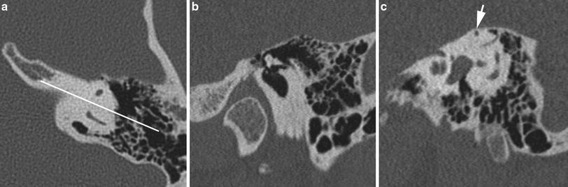
Fig. 2
Stenvers’ Plane. Single Oblique Reconstruction perpendicular to the Plane of the SSC (Long Axis of the Temporal Bone). a Axial reference image demonstrating plane of reconstruction (white line) perpendicular to the plane of the SSC. b Multiplanar reconstruction in the Stenvers Plane through the middle ear at the level of the incudomalleolar joint. c Stenvers MPR through the inner ear at the level of the apex of the SSC (white arrow)
Additional targeted MPRs can be employed to investigate particular anatomic structures in a more detailed manor. Most of the targeted MPRs employed in this chapter are slight modifications of standard imaging planes used in temporal bone polytomography (sagittal, coronal, Pöschl, Stenvers planes). It is often useful to have the capability of performing reconstructions in two independent oblique planes (double oblique reconstruction) in order to optimally profile specific anatomic structures, in particular the auditory ossicles.
1 Technical Considerations
In general, the higher the number of detector rows employed by a particular MDCT scanner, the more isotropic the voxel size and the greater the resolution of the MPRs. All MPR images included in this chapter were reconstructed from volumetric data sets acquired on a 64-slice MDCT yielding a voxel size of 0.4 mm3. Supplemental short axis (Pöschl) and long axis (Stenvers) MPR volumes can be routinely reconstructed at the CT console using the previously mentioned anatomic landmarks at the request of the interpreting radiologist. Any additional targeted MPRs are reconstructed from the volumetric data set at a separate workstation, preferably one that is capable of producing MPRs using at least two independent planes of obliquity.
2 Middle Ear
Long axis MPRs and maximum intensity projections (MIPs) of the ossicles can be useful in assessment of ossicular integrity (Bin et al. 2008). In the clinical setting of conductive hearing loss. Chronic inflammatory disease with or without cholesteatoma, temporal bone fracture, or congenital anomalies such as aural atresia or microtia can produce various ossicular pathologies to include fixation, erosions, and disarticulations. Since the standard axial and coronal orientations do not display the ossicles to best advantage, the integrity of the ossicular chain can be confirmed with long axis MPRs of each individual ossicle (Figs. 3, 4, 5). Occasionally MPRs in the plane of the short axis of the stapes can be useful, particularly in the setting of stapes footplate fixation secondary to early otospongiotic plaque along the anterior edge of the oval window in the vicinity of the fissula ante fenestram (Fig. 6).
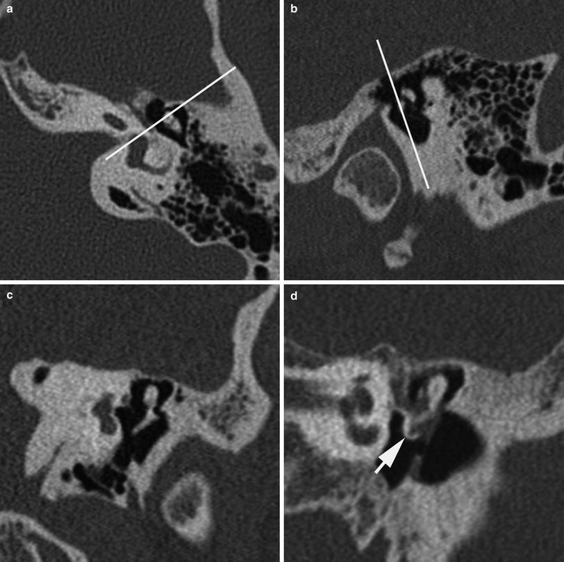
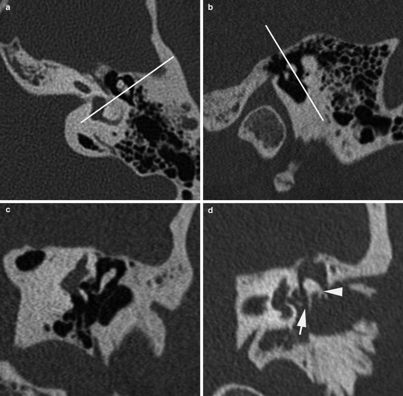
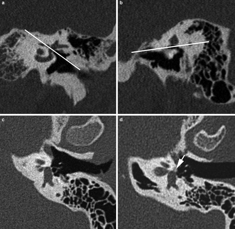
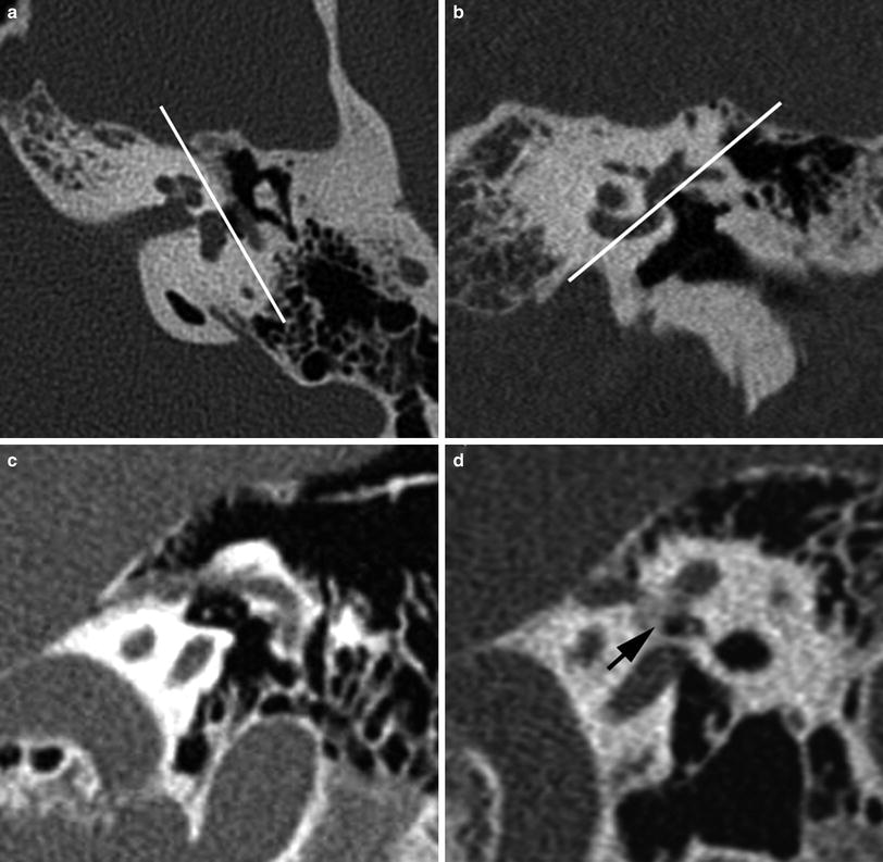
Fig. 3
Long Axis Coronal Oblique Reconstruction of the Malleus. a Axial and b sagittal references images demonstrating multiplanar reconstruction plane required for the coronal long axis view of the malleus. c Multiplanar reconstruction (MPR) of the malleus from MDCT data set. d MPR demonstrates fixation of the manubrium of malleus to calcified chronic inflammatory tissue (tympanosclerosis) (arrow) in this patient with conductive hearing loss. [Reprinted with permission from Springer (Lane & Witte, Temporal Bone: An Imaging Atlas, Springer)]
Fig. 4
Long Axis Coronal Oblique Reconstruction of the Incus. a axial and b sagittal references images demonstrating multiplanar reconstruction planes required for the coronal long axis view of the incus. c multiplanar reconstruction (MPR) of the incus from MDCT data set. d MPR demonstrates incudostapedial disarticulation (arrow) with rotational dislocation of the incus. Note short process is rotated laterally from its normal posterior orientation (arrowhead) in this patient with acute longitudinal fracture of the left temporal bone. [Reprinted with permission from Springer (Lane & Witte, Temporal Bone: An Imaging Atlas, Springer)]
Fig. 5
Long Axis Axial Oblique Reconstruction of the Stapes. a Coronal and b sagittal references images demonstrating multiplanar reconstruction plane required for the long axis oblique axial view of the stapes. c multiplanar reconstruction (MRP) of the normal stapes. d MPR demonstrates fixation of the footplate and anterior crus of the stapes secondary to fenestral otospongiosis. Note stapes seen in long axis with demineralized, otospongiotic bone arising from the region of the fissula ante fenestram and encasing the anterior edge of the footplate and anterior crus (arrow). [Reprinted with permission from Springer (Lane & Witte, Temporal Bone: An Imaging Atlas, Springer)]
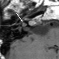
Fig. 6
Short Axis Sagittal Oblique Reconstruction of the Stapes. a Axial and b coronal references images demonstrating multiplanar reconstruction planes required for the short axis oblique sagittal view of the stapes. c multiplanar reconstruction (MPR) of the stapes demonstrating anterior and posterior crura in the oval window niche. d MPR demonstrates fixation of the footplate and anterior crus of the stapes secondary to fenestral otospongiosis. Note stapes seen in short axis with demineralized, otospongiotic bone arising from the region of the fissula ante fenestram and encasing the anterior crus (black arrow). Note that the posterior crus is not involved. [Reprinted with permission from Springer (Lane & Witte, Temporal Bone: An Imaging Atlas, Springer)]
An oblique sagittal MPR can be employed to profile the incudomalleolar joint and at the same time inspect the integrity of the manubrium of the malleus and long process of the incus, recreating the old “Molar Tooth” tomographic appearance (Fig. 7). Since the malleus and incus differ slightly in their lateral to medial angulation, a 2–3 mm MIP in the oblique sagittal plane can be quite helpful in producing a more complete assessment of these ossicles (Fig. 7c). This perspective can be useful in confirming any suspicion of malleoincudal luxation seen on axial or coronal images in patients with conductive hearing loss following trauma (Fig. 7e, f). Erosions of the long process of the incus, a relatively common complication of middle ear inflammatory disease, can be nicely demonstrated in this plane with both MPR and MIP images (Fig. 7g, h). Additionally, Pöschl plane reconstructions of the ossicles provide an ideal depiction of both the incudomalleolar and incudostapedial articulations (Fig. 8). Pöschl MPR and MIP images can be useful in confirming incudostapedial disarticulation following trauma or as a result of chronic inflammatory disease (Fig. 8d).
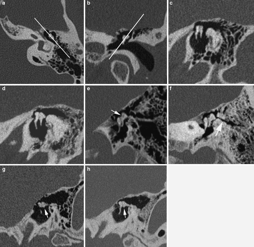
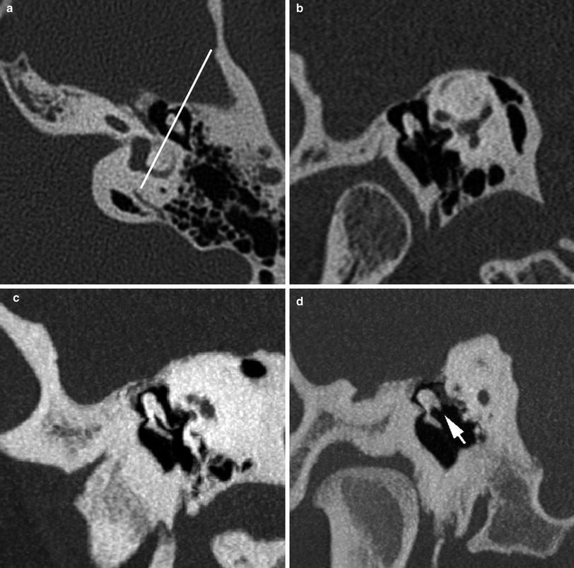
Fig. 7
Long Axis Sagittal Oblique Reconstruction of the Malleus and Incus (Molar Tooth View). a Axial and b coronal references images demonstrating multiplanar reconstruction plane required to view the sagittal long axis of the malleus and incus. c multiplanar reconstruction (MPR) of the malleus and incus from multidetector CT data set. Note normal relationship of malleus anterior to incus with mallear head articulating with articular facet of Incus. d Maximum intensity projection (MIP) (2 mm slab) of malleus and incus. e MPR of disarticulated malleus and incus in patient with conductive hearing loss following remote trauma. Note ‘empty’ articular facet of the incus (arrow) with inferiorly displaced malleus relative to incus. f MIP (2 mm slab) of disarticulated malleus and incus. Note ununited fracture plane (arrow). g MPR demonstrates erosion of the long process of the incus (arrow) on MPR and, h on MIP (2 mm slab). [Reprinted with permission from Springer (Lane & Witte, Temporal Bone: An Imaging Atlas, Springer)]
Fig. 8


Pöschl’s plane Reconstruction of the incudomallear and incudostapedial articulations. a Axial reference image demonstrating multiplanar reconstruction in the plane of the short axis of the temporal bone required for this reconstruction. b Multiplanar reconstruction (MPR) of the ossicles in Pöschl’s plane. c Maximum intensity projection (MIP) (2 mm slab) of normal ossicular articulations. d MIP (2 mm slab) of eroded long process of the incus (arrow) with disarticulation of the incudostapedial joint in a patient with conductive hearing loss. [Reprinted with permission from Springer (Lane & Witte, Temporal Bone: An Imaging Atlas, Springer)]
Related posts:


Stay updated, free articles. Join our Telegram channel
Full access? Get Clinical Tree
