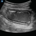
Figure 9.1
Right kidney: The right kidney is visualized next to the liver in long axis

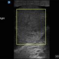
Figure 9.2
Right kidney in short axis: In short axis, the kidney will appear more rounded

Figure 9.3
Left kidney: The left kidney is visualized next to the spleen in long axis

Figure 9.4
Left kidney in short axis: The left kidney in short axis next to the spleen

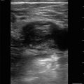
Figure 9.5
Bladder in transverse: Cross-sectional view of the bladder

Figure 9.6
Bladder in sagittal: Sagittal view of the bladder. Note the posterior acoustic enhancement that is normal for fluid filled structures

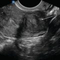
Figure 9.7
Uterus posterior to bladder: In females, the uterus is located directly posterior to the bladder (arrow)

Figure 9.8
Prostate posterior to bladder: In males, the prostate is located posterior to the bladder (arrow)

Figure 9.9
Normal bladder wall thickness: Bladder wall thickness measured at 0.15 cm. Normal bladder wall thickness is less than 5 mm when empty and less than 3 mm when full

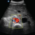
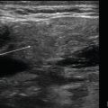
Figure 9.10
Ureteral flow jets with color Doppler: In a transverse plane, ureteral flow jets representing urine entering the bladder can be seen. Color Doppler can enhance the detection of ureteral flow jets

Figure 9.11
Bladder volume measurement in transverse: In a transverse plane, measure across the bladder to obtain length (L) and from anterior to posterior to obtain width (W)

Figure 9.12
Bladder volume measurement in sagittal: In sagittal orientation, measure superior to inferior to obtain the height (H) measurement
Renal Pathology
- (a)
Hydronephrosis
Dilation of the renal collecting system due to intrinsic or extrinsic obstruction:
Common etiologies include large ureteral stone, enlarged prostrate, bladder mass, and extrinsic compression of collecting system.
Appears as an anechoic fluid collection within the renal sinus.
Hydronephrosis can be mild, moderate, or severe:
- (b)
Hydroureter
Dilated ureter
Can be seen with severe hydronephrosis
Figure 9.16—Hydroureter
Video 9.12—HydroureterRelated posts:

Stay updated, free articles. Join our Telegram channel

Full access? Get Clinical Tree