Outline
Imaging Techniques, 1046
Anatomy and Physiology, 1046
Congenital Anomalies, 1048
Leiomyomas, 1051
Definition and Epidemiology, 1051
Clinical Aspects, 1051
Pathophysiology, 1052
Treatment, 1053
Diagnosis, 1053
Imaging Characteristics, 1053
Adenomyosis, 1056
Definition and Epidemiology, 1056
Clinical Aspects, 1056
Pathophysiology, 1056
Diagnosis, 1056
Imaging Characteristics, 1056
Endometriosis, 1056
Cervical Cancer, 1057
Epidemiology and Clinical Presentation, 1057
Histopathology, 1059
Diagnosis, 1060
Treatment, 1060
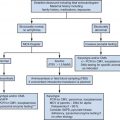
Clinical Staging, 1060
Imaging, 1060
Endometrial Cancer, 1063
Epidemiology and Clinical Presentation, 1063
Histopathology, 1063
Diagnosis, 1063
Treatment, 1063
Staging, 1064
Imaging, 1064
Adnexal Masses, 1065
Epidemiology and Clinical Significance, 1065
Imaging Diagnosis and the Role of Magnetic Resonance Imaging, 1066
Differentiation of Benign and Malignant Lesions, 1067
Specific Diagnosis Based on Magnetic Resonance Imaging Findings, 1067
Conclusion, 1070
Summary of Key Points
- •
Magnetic resonance imaging (MRI) is a problem-solving tool and has greater accuracy and specificity for characterization of gynecologic masses than sonography or computed tomography (CT), but should generally be performed after pelvic sonography.
- •
High resolutionT2-weighted sequences are the mainstay images for evaluation of nonobstetric gynecologic disorders.
- •
T1-weighted imaging must be performed with and without fat suppression if characterization of fat or blood products is required.
- •
The uterine corpus is best assessed in the parasagittal plane.
- •
The cervix is best assessed in the axial plane.
- •
MRI is the method of choice for evaluation of congenital uterine anomalies, including associated renal anomalies.
- •
Water-based gel instilled into the vagina is helpful in cervical and vaginal cancer assessment.
- •
Gadolinium is helpful in defining the extent and vascularity of tumors.
- •
Many tumors demonstrate restricted diffusion.
- •
Staging of gynecologic malignancy is based on the International Federation of Gynecology and Obstetrics (FIGO) system, and cross-sectional imaging is helpful in preoperative planning.
This chapter describes the main indications for MRI in the evaluation of nonobstetric gynecologic disorders. In the first two sections, normal anatomy of the female pelvis and common congenital anomalies are discussed. In subsequent sections, the use of MRI in the setting of common benign diseases, namely leiomyomas, adenomyosis, and endometriosis is outlined. Finally, the role of MRI in patients with malignant neoplasms of the reproductive organs is discussed. The main clinical and epidemiologic aspects of gynecologic disease are presented, in addition to the imaging features, in order to emphasize a practical clinical approach to patient management.
Imaging Techniques
Although a detailed discussion of imaging techniques and protocols used in MRI is beyond the scope of this chapter, some of the most important basic concepts with regard to MRI techniques are included to aid in interpretation of images presented herein, targeting those readers unfamiliar with MRI.
T2-weighted images provide the foundation for MRI of the female pelvis. These images are used for evaluation of normal anatomy and detection of most uterine and adnexal abnormalities. T1-weighted images, especially when used in conjunction with fat suppression, are helpful in characterizing the content of adnexal lesions, namely in the identification of blood and fat. When used after the administration of intravenous gadolinium, T1-weighted images also assist in the characterization of some benign and malignant tumors. As a general rule, on T1-weighted images, fat-containing lesions are bright (high signal intensity), whereas water, such as urine in the bladder, is dark (low signal intensity). However, water is bright on T2-weighted images. Therefore, tissues with higher amounts of water, such as tumors and cysts, have high signal intensity on T2-weighted images. Water-based gel instilled into the vagina can be used to distend the vaginal lumen and outline the cervical contour, but this technique is not widely used. Gadolinium-based contrast agents are most commonly used as intravenous contrast media in MRI. Tissues that are infused with gadolinium increase in signal intensity on T1-weighted images when compared with images acquired before contrast administration. Hence, gadolinium is used to increase the contrast between normal and abnormal tissues in the body and to detect vascularity of tissues. For instance, many neoplastic processes enhance more avidly than adjacent normal tissues and thus become more easily visible. Newer techniques such as dynamic multiphase contrast-enhanced MRI (DCE-MRI) and diffusion-weighted imaging (DWI) provide functional information. In DCE-MRI, images are acquired at multiple times following injection of contrast agent, allowing assessment of tissue vascularity and enhancement patterns. DWI does not use intravenous contrast, but displays contrast between tissues based on different rates of diffusion of water molecules. Many malignant tumors are bright on DWI.
Anatomy and Physiology
Although it is not within the scope of this chapter to fully discuss the normal anatomy of the female pelvis, a few words regarding specific features on MRI are important. A basic concept that must be remembered is that the patient’s hormonal status affects the imaging features of the female reproductive organs, irrespective of the imaging modality.
Uterus
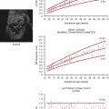
The uterus typically measures from 6 to 9 cm in length in women of reproductive age; the cervix accounts for approximately 30% to 50% of the total uterine length in this age group, but is proportionally larger before the menarche. Uterine zonal anatomy is best visualized using T2-weighted images, typically acquired in the axial and sagittal planes, and may be described by considering two separate regions, the cervix and the uterine body.
The cervical anatomy is divided into three zones on T2-weighted sequences: the endocervical mucosa centrally, which demonstrates high signal intensity as a result of the presence of mucus within glands; the cervical stroma, which demonstrates low signal intensity owing to the presence of fibrous connective tissue; and the peripherally located smooth muscle, which demonstrates intermediate signal intensity ( Fig. 36-1 ). Varying amounts of mucus may be seen within the endocervical canal, which has a very high signal intensity similar to that of fluid.
The zonal anatomy of the body of the uterus also consists of three distinct regions on T2-weighted sequences: centrally, the endometrium demonstrates very high signal intensity; subjacent to the endometrium, the junctional zone, which represents the innermost layer of the myometrium, demonstrates low signal intensity; and the outer myometrium, which demonstrates intermediate signal intensity ( Fig. 36-2 ). At its inferior aspect, the junctional zone is continuous with the low signal intensity fibrous cervical stroma.
Identification of the junctional zone is extremely important when evaluating the uterine magnetic resonance zonal anatomy. Studies have shown that the appearance of the zonal anatomy on MRI is influenced by female hormones. Normally, the junctional zone should not measure more than 11 mm in thickness in women of reproductive age. In postmenopausal women, women taking oral contraceptives, or in girls before menarche, the zonal anatomy is frequently indistinct and the junctional zone may be very thin.
The MRI appearance of the endometrium is also hormonally influenced and varies with the menstrual cycle. The endometrium becomes progressively thicker between the early follicular (proliferative) phase and ovulation, increasing from 1 to 3 mm in thickness to an average of 8 to 9 mm. The endometrium should not measure more than 15 mm in thickness in women of menstrual age, and typically it measures 11 mm or less in postmenopausal women who are not on hormonal replacement therapy. The endometrium is highest in signal intensity during the luteal (secretory) phase. It is imperative to recognize that the evaluation of endometrium thickness using T2-weighted images may result in overestimation because fluid within the endometrial cavity is also high in signal intensity on T2-weighted sequences ( Fig. 36-3 ).
Unlike the uterine corpus, the cervical zonal anatomy does not demonstrate significant changes during the menstrual cycle. In addition, because the cervix is composed largely of fibrous elastic tissue, enhancement of the cervix following the administration of intravenous gadolinium differs from enhancement of the uterine corpus. The uterine corpus is a very vascular structure when compared to the cervix and enhances earlier and more avidly than the cervix ( Fig. 36-4 ). This is important because this difference in enhancement pattern may give the appearance of a pseudomass and lead to a misdiagnosis of cervical cancer.
Ovaries
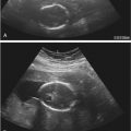
The normal ovaries demonstrate homogeneous intermediate T1-weighted signal intensity; however, on T2-weighted sequences, an outer high signal intensity cortex and a hypointense central medulla will be observed. The ovaries measure approximately 2 × 2 × 3 cm; however, their size fluctuates according to hormonal status, largely because of follicle production and enlargement during the menstrual cycle. The ovaries are most readily identified in women of reproductive age by the presence of such follicles, which appear as small simple cysts of high signal intensity on T2-weighted sequences and low signal intensity on T1-weighted sequences ( Fig. 36-5 ). Primordial follicles are usually smaller, measuring up to 9 mm in diameter, whereas stimulated graafian follicles may reach up to 3 to 5 cm. Other features of these physiologic cysts seen on T2-weighted sequences include imperceptible or thin walls (less than 3 mm) and absence of mural nodules (see Fig. 36-5 ). The number of ovarian follicles decreases following menopause in women who are not taking hormonal replacement therapy. However, simple ovarian cysts are a common incidental finding in postmenopausal women, seen in 14% of women over 55 years of age, and may resolve or remain stable. Ovarian stroma enhances after intravenous contrast agent administration and the use of fat-suppressed sequences helps to identify the ovaries when follicles are not visible. The normal fallopian tubes are not visualized on MRI.
Congenital Anomalies
MRI is the imaging method of choice for evaluation of congenital anomalies of the female reproductive tract. Advantages of MRI include noninvasiveness, lack of ionizing radiation, multiplanar capability enabling visualization of the fundal contour of the uterus, and excellent soft tissue characterization. The use of MRI may reduce the number of invasive procedures and related costs by guiding management decisions.
Müllerian Duct Anomalies
The müllerian ducts are paired embryologic structures that normally fuse between the 6th and 11th weeks of gestation, forming the uterus, fallopian tubes, cervix, and most of the upper vagina. However, in 0.1% to 10.0% of the general population, either fusion fails to occur or the ducts do not develop normally, resulting in one of the multiple types of anomalies. Although müllerian duct anomalies may be asymptomatic depending upon the exact type, they may be associated with primary amenorrhea, infertility, obstetric complications, and endometriosis. It is estimated that as many as 25% of women who present with infertility and miscarriage have müllerian duct anomalies. In addition, 30% to 50% of these women have concurrent renal anomalies, which include renal agenesis, ectopia, fusion, malrotation, and duplication. Patients suspected of having müllerian duct anomalies are often initially investigated with transvaginal sonography, because it is widely available, safe, and of relatively low cost. Although MRI is considered the reference standard for the evaluation of müllerian duct anomalies with a diagnostic accuracy of almost 100%, it is frequently reserved for indeterminate cases or cases in which further information is needed. T2-weighted sequences are the most useful because identification of the zonal anatomy is necessary for complete evaluation and analysis. In addition, the study must include an oblique coronal and axial plane to ensure adequate visualization of the uterine fundal contour.
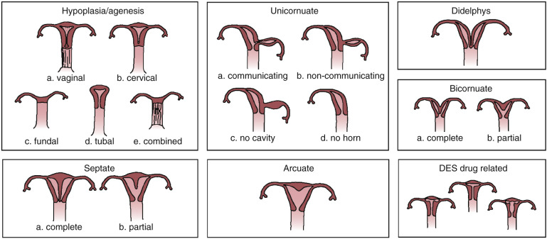
Although there are many different classifications of müllerian duct anomalies, the American Society for Reproductive Medicine classification is perhaps the most widely used and the best known ( Fig. 36-6 ).
Agenesis/Hypoplasia
The anomalies within this group result from bilateral nondevelopment or arrested development of the müllerian ducts. Agenesis of the upper vagina and uterus is the most common anomaly in this group and part of the spectrum of the Mayer-Rokitansky-Küster-Hauser syndrome. Patients with this syndrome may present with an atrophic uterus, however, and vaginal abnormalities can range from hypoplasia to agenesis. Urinary system malformations are sometimes associated with the Mayer-Rokitansky-Küster-Hauser syndrome. The second most common anomaly in this class is segmental vaginal agenesis. Isolated agenesis or hypoplasia of the uterus is very rare. In complete uterine agenesis, MRI demonstrates only a short blind-ending vagina, whereas in cases of uterine hypoplasia, the uterus is small and the intercornual diameter of the endometrial cavity is reduced, measuring less than 2.0 cm.
Unicornuate Uterus
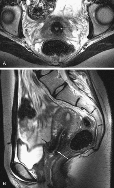
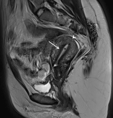
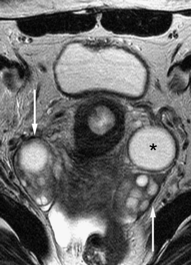
A unicornuate uterus is the consequence of complete arrest or partial development of one müllerian duct in utero. In 90% of cases, partial development of the duct occurs, with an ensuing rudimentary horn, which may or may not contain functioning endometrium. The rudimentary horn may be obstructed or may communicate with the main uterine cavity. Clinical associations include very high rates of spontaneous miscarriage, obstetric complications, and endometriosis. A unicornuate uterus will have normal zonal anatomy and has a characteristic banana-shaped configuration. A rudimentary horn can be seen as an adjacent soft tissue mass and may be distinguished from a tumor by virtue of signal intensity characteristics that are similar to myometrium on T2-weighted images. An obstructed (noncommunicating) rudimentary horn may become enlarged as a result of distention from blood products, identified as high signal intensity central material on T1-weighted sequences.
Uterus Didelphys
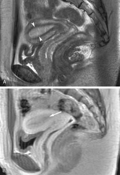
Uterus didelphys arises through complete failure of müllerian duct fusion in utero and is characterized by the presence of two separate uteri and cervices with a vertical septum in the upper vagina. Very rarely, two completely separated vaginas and vaginal orifices may be present. Patients are usually asymptomatic, but hematometrocolpos may occur in the presence of an obstructing transverse hemivaginal septum. Clinical complications are similar to those associated with a unicornuate uterus. MRI may be used to readily diagnose uterus didelphys by demonstration of two widely separated uterine horns and two cervices ( Fig. 36-7 ). The zonal anatomy is preserved. Vaginal septa may not always be identified with MRI, and physical examination may be necessary for confirmation and differentiation from a bicornuate bicollis uterus.

Bicornuate Uterus
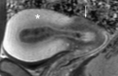
This anomaly is the result of partial failure of fusion of the müllerian ducts. It is characterized by an increased intercornual distance, reportedly measuring wider than 4 cm; a concave external contour of the fundus, measuring more than 1 cm in depth; and a fibrous-muscular septum bisecting the endometrial cavity. The septum may be incomplete, partially dividing the uterine cavity (bicornuate unicollis [ Fig. 36-8 ]), or complete, extending all the way to the external cervical os (bicornuate bicollis). The vagina develops normally. Images obtained in the oblique coronal plane, along the long axis of the uterus, are useful in assessing the fundal contour. The dividing septum often exhibits signal characteristics similar to myometrium, but may be of low signal intensity inferiorly when fibrous tissue is present.

Septate Uterus
Septate uterus is the most common müllerian duct anomaly and is thought to result from failure of resorption of a fibrous septum in later stages of development. The result of such failure of resorption is a characteristically small endometrial cavity with a dividing septum, which may be partial or may extend to the external cervical os. Reproductive complications are greatest in this class of müllerian duct anomaly and are attributed to difficulties of implantation. Identification of a convex, flat, or minimally indented fundal contour without a fundal cleft on MRI obtained in the coronal plane ( Fig. 36-9 ) is decisive in distinguishing a septate from a bicornuate uterus. Other diagnostic MRI features include an intercornual distance of less than 4 cm (i.e., not increased) and a thin, dividing, often fibrous septum typically of low T1- and T2-weighted signal intensity, although the septum may have a signal intensity similar to that of myometrium if myometrial smooth muscle is the primary component of the septum. The septum may be partial or complete.

Arcuate Uterus
This anomaly is recognized by mild indentation of the myometrium into the fundal aspect of the endometrium, attributed to minimally incomplete resorption of the uterovaginal septum. It is classified separately because it is a benign variant without clinical significance. On MRI, the arcuate uterus is typically of normal size with a single endometrial cavity that is minimally indented superiorly by the small nonresorbed septal remnant and may be associated with a small fundal cleft less than 1.0 cm in depth.
Diethylstilbestrol Exposure
Diethylstilbestrol is a synthetic estrogen that was historically prescribed to prevent miscarriages until 1971. It is estimated that approximately two thirds of patients exposed to this drug while in utero have uterine anomalies. The most common of these anomalies is the classic hypoplastic T-shaped uterus, but other deformities of the uterine horns and endometrial cavity have been reported.
Vaginal Anomalies
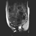
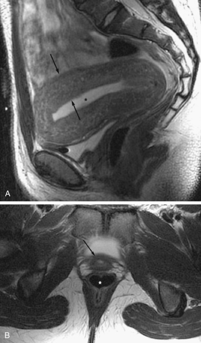
The lower third of the vagina originates from the urogenital sinus rather than the müllerian ducts and is developmentally distinct. Absence of the distal or lower third of the vagina or the presence of a transverse membrane (mimicking an imperforate hymen) results from failure of urogenital sinus development. In both eventualities, the vaginal obstruction causes the vagina and uterus to distend with fluid, referred to as hematometrocolpos when blood products are identified. The upper vagina demonstrates very thin walls, whereas the uterus may have either thin or thick myometrium ( Fig. 36-10 ). Hematometrocolpos is characterized as a heterogeneous collection with high T1- and intermediate or low T2-weighted signal intensity.
Ovarian Anomalies
The ovaries arise from primitive germ cells that migrate to the embryonic gonadal ridge and subsequently descend into the pelvis. Congenital anomalies of the ovaries are typically associated with chromosomal disorders and include gonadal dysgenesis, commonly occurring with Turner syndrome (45,X karyotype) and Turner syndrome mosaicism (45,X/46XX karyotype). The ovaries of patients with gonadal dysgenesis are absent or seen as fibrous streaks. Fat-suppressed T2-weighted and gadolinium-enhanced T1-weighted sequences are most helpful in the identification of the ovaries.
Leiomyomas
Definition and Epidemiology
Leiomyomas are benign tumors arising from uterine smooth muscle cells and occur in more than 80% of African-American women and in approximately 70% of Caucasian women by age 50 years. African-American women are diagnosed with leiomyomas at earlier ages, are more likely to be symptomatic, and are more likely to undergo myomectomies and hysterectomies compared to Caucasian women. Leiomyomas have a significant impact on public health, accounting for at least one third of the 600,000 hysterectomies performed each year in the United States.
Clinical Aspects
Most patients with leiomyomas are asymptomatic. Clinical symptoms associated with leiomyomas include dysfunctional uterine bleeding, pelvic pressure, and pelvic pain. In addition, there is an association with impaired reproductive function, which may be related to impaired implantation, tubal obstruction, and an increased rate of miscarriage as well as preterm labor. Approximately 90% of leiomyomas occur in the uterus, whereas 5% to 10% are cervical in location. Leiomyomas can enlarge during pregnancy, and cervical leiomyomas may cause dystocia. A minority of leiomyomas occur in the round and broad ligaments ( Fig. 36-11 ) or adnexa.

Location of the leiomyoma often predicates the symptoms. Uterine leiomyomas are classified as submucosal, intramural, or subserosal ( Fig. 36-12 ). Approximately 5% of leiomyomas are submucosal in location ( Fig. 36-13 ). Submucosal leiomyomas typically present with menometrorrhagia. In addition, a submucosal leiomyoma is more likely to be associated with infertility, dysfunctional uterine bleeding, dysmenorrhea, and anemia. Submucosal leiomyomas are often effectively treated with hysteroscopic resection, depending upon size and intracavitary extent of the mass. Submucosal leiomyomas are distinguished from large intramural leiomyomas based on identification of the lesion’s epicenter within the endometrial cavity rather than within the myometrium, and less than 180-degree encirclement by surrounding myometrium.


Subserosal leiomyomas account for approximately 10% to 20% of uterine leiomyomas, are located just under the uterine serosa, and may be pedunculated or sessile ( Figs. 36-13 and 36-14 ). Symptoms are associated with large size owing to mass effect or torsion of a pedunculated lesion. The bridging vascular sign (vessels originating from the uterus and feeding a pelvic mass) is seen in approximately 77% of exophytic leiomyomas and is helpful in differentiating these tumors from solid adnexal masses. Conversely, an ovarian vascular pedicle (identified by the presence of a gonadal vein) may be helpful in establishing that a mass is ovarian in origin. A pedicle is seen in 92% of ovarian masses and 13% of pedunculated subserosal leiomyomas.

Pathophysiology
The pathophysiology of leiomyomas is unknown; however, genetic predisposition and hormonal status are involved in tumor development and growth. Responsiveness of leiomyomas to hormonal stimulation frequently results in enlargement during pregnancy and involution following menopause. Various types of benign degeneration are common, particularly in large leiomyomas, and include hyaline (most common and present in at least 60% of leiomyomas), cystic, myxoid, fatty, and hemorrhagic degeneration ( Fig. 36-15 ). Imaging-based differentiation between the types of degeneration is usually not possible; however, certain features may favor one or another type. Degeneration within a leiomyoma may make the sonographic diagnosis difficult as a degenerated leiomyoma may have cystic components and, thus, may mimic the sonographic appearance of a complex ovarian mass. In such cases, MRI may be used for diagnosis.

Knowledge of atypical locations and presentations, such as benign metastasizing leiomyomas, intravenous leiomyomas, diffuse leiomyomatosis, and peritoneal disseminated leiomyomatosis, is important. Although these uncommon presentations may suggest a malignant process, they are essentially a variation in the growth pattern of this benign lesion. Uterine leiomyosarcomas have been reported to occur in association with leiomyomas. However, genetic studies suggest that these are two different entities arising from separate pathways. Malignant degeneration of a benign leiomyoma into a leiomyosarcoma is considered by most pathologists to be extremely rare, if it occurs at all. Sometimes, the distinction between a leiomyoma and a leiomyosarcoma cannot be reliably made based on MRI characteristics of the primary lesion; however, rapid growth, irregular borders, evidence of extrauterine extension, lymphadenopathy, and metastases are, to different degrees, suggestive of malignancy ( Fig. 36-16 ).

Treatment
The preferred treatment for leiomyomas depends upon their size and location, as well as clinical presentation. The most common therapeutic options are hysterectomy, myomectomy (transabdominal, laparoscopic, or hysteroscopic), myolysis, and uterine artery embolization (UAE). Hysterectomy is usually performed in women who have completed childbearing and do not wish to preserve the uterus. Myomectomy is often the choice for those patients who wish to preserve the uterus. In these cases, an abdominal approach is used for multiple large leiomyomas; the laparoscopic approach is appropriate for resection of subserosal and pedunculated lesions, whereas the hysteroscopic technique is preferred for patients with submucosal lesions that are greater than 50% intracavitary. UAE is a newer, safe, and effective technique with the potential to become the treatment of choice for symptomatic uterine leiomyomas. Contraindications to UAE include active pelvic infection, renal insufficiency, and contrast agent allergy. The reported principal advantage of UAE in appropriate candidates is lower morbidity compared to more invasive surgical techniques.
Diagnosis
The most common indication for MRI of symptomatic patients is to determine the size and location of leiomyomas and to assess for the presence of necrosis or hemorrhage in lesions that appear worrisome on ultrasound imaging, as such factors are essential for guiding appropriate patient management. The initial diagnosis of leiomyoma is typically confirmed and then monitored, as clinically indicated, by pelvic sonography. Ultrasound evaluation may be limited in a number of situations, such as retroverted or retroflexed uterus; uterine or leiomyoma enlargement beyond the sonographic field of view, precluding complete visualization; pedunculated subserosal leiomyomas, which can be difficult to distinguish from adnexal masses; and atypical imaging features. In such cases, MRI is useful as a problem-solving tool and for preprocedural assessment when limited or conservative treatment is planned. Although MRI and sonography have similar overall sensitivity (99% for each) for detection, sonography has a lower reported specificity than MRI (86% and 91%, respectively). Furthermore, MRI is better for determining the total number of lesions. The advantages of MRI over sonography in this application are primarily due to the larger field of view and to significantly improved soft tissue characterization. An additional clinically important benefit of MRI is the ability to readily differenetiate leiomyomas from focal adenomyosis, as these two entities are managed differently if uterine preservation is desired. Finally, MRI has a role in predicting and monitoring treatment response, especially in patients treated with UAE. Pretreatment imaging features predictive of a good response to UAE include submucosal location, small size, avid contrast enhancement (vascularity), and high T2-weighted signal intensity. Conversely, the presence of hemorrhagic degeneration or lack of contrast enhancement before embolization often predicts a poor response. Successful UAE results in hemorrhagic infarction and ultimate reduction in size of both the uterus and any leiomyomas.
Imaging Characteristics
On MRI, a simple nondegenerating leiomyoma is depicted as a well-circumscribed, round mass with low signal intensity on both T1- and T2-weighted sequences (see Fig. 36-15 ). Intramural leiomyomas may demonstrate a high signal intensity pseudocapsule on T2-weighted sequences, which has been attributed to edema and vascular or lymphatic congestion within the surrounding myometrium ( Fig. 36-17 ). Calcifications may be seen as areas of signal void but are generally not well visualized with MRI. Nondegenerating leiomyomas typically show marked enhancement after gadolinium administration, but the use of contrast material is required only for the purpose of determining lesion vascularity when considering UAE (see Fig. 36-15 ).

As mentioned previously, imaging characteristics of degeneration are nonspecific, but some MRI features may suggest specific types. Leiomyomas with cystic degeneration present with very high signal intensity on T2-weighted sequences ( Fig. 36-18 ). Heterogeneous low T2-weighted signal intensity and a cobblestone or whorled appearance in a leiomyoma is usually associated with hyaline degeneration. Hemorrhagic degeneration commonly occurs following UAE or as a sequela of rapid lesion enlargement during pregnancy. Hemorrhagic degeneration characteristically manifests as high signal intensity on T1-weighted sequences because of the high proteinaceous content of blood or the presence of methemoglobin (see Fig. 36-15 ). A rim of low signal intensity on T2-weighted sequences and high signal intensity on T1-weighted sequences may be present and is thought to represent obstructed veins around the lesion. Myxoid degeneration is associated with multiple areas of cystic change and very high T2-weighted signal intensity. No specific MRI features have been described that reliably differentiate a leiomyoma from a leiomyosarcoma. However, an irregular, infiltrative, ill-defined border; extrauterine extension; and associated pelvic lymphadenopathy are worrisome features for malignancy, as noted earlier (see Fig. 36-16 ).

Adenomyosis
Definition and Epidemiology
Adenomyosis refers to the presence of intramyometrial endometrial mucosa (both glands and stroma) surrounded by hypertrophied myometrium. Typically, adenomyosis is diagnosed in a multiparous woman in the fourth or fifth decade of life. Although common, the exact prevalence of adenomyosis is difficult to establish. Autopsy and clinical studies on prevalence vary, with a range of 20% to 67% in autopsy studies and 10% to 88% in clinical studies ; this wide range of reported prevalence is likely a reflection of several factors, including inconsistent pathologic definition of adenomyosis, different uterine specimen processing protocols, and varied patient inclusion criteria.
Clinical Aspects
The classic clinical scenario is that of a patient who presents with menorrhagia, dysmenorrhea, and metrorrhagia accompanied by an enlarged, smooth-contoured soft uterus that may be tender to palpation. However, one third of patients with adenomyosis are asymptomatic. Both the frequency and severity of symptoms appear to correlate with the extent and depth of myometrial involvement.
Pathophysiology
Adenomyosis causes globular enlargement of the uterus with cystic areas, some filled with degraded red blood cells. Adenomyosis may be described as focal, diffuse, or a combination, depending on its distribution within the myometrium. Irrespective of presentation, these lesions have ill-defined margins, unlike leiomyomas, which are sharply marginated with a smooth, regular border.
There are three general hypotheses that attempt to explain the development of adenomyosis. Two hypotheses suggest a process of invagination of the basalis layer of the endometrial mucosa, either between muscle fibers or along the intramyometrial lymphatic system. The third theory suggests that adenomyosis develops as a result of metaplasia from de novo ectopic intramyometrial endometrial tissue. In up to 80% of cases, there are additional uterine pathologic findings, most commonly leiomyomas, but also endometrial polyps, endometrial hyperplasia, and endometrial adenocarcinoma.
Diagnosis
Although presentation may strongly suggest adenomyosis, clinical diagnosis is not accurate, and imaging is often used prior to treatment selection.
Imaging Characteristics
Transvaginal sonography is generally the first choice imaging modality in symptomatic patients with adenomyosis. It is widely available, safe, and low cost with good accuracy, similar to that reported for MRI. Reported sensitivity of transvaginal sonography ranges from 57% to 89% and specificity from 65% to 98%. However, ultrasound evaluation is somewhat operator dependent, with the best results obtained by experienced examiners. In addition, the accuracy of sonography decreases in patients who present with large uteri and concurrent leiomyomas. The reported sensitivity and specificity of MRI range from 70% to 86% and 86% to 93%, respectively. MRI is generally recommended if sonographic results are equivocal or when additional information is needed, especially for treatment planning.
The diagnosis of adenomyosis on MRI is based on findings identified on T2-weighted sequences. Classically, adenomyosis appears on MRI as focal or diffuse widening of the junctional zone (inner myometrium) or as an ill-defined, hypointense, myometrial mass (see Figs. 36-14 and 36-17 ). The normal junctional zone in premenopausal women appears as a band of low signal intensity between the endometrium and outer myometrium. Based on the study by Reinhold and associates, identification of a junctional zone wider than 12 mm is diagnostic of adenomyosis, whereas a width of 8 mm or less reliably excludes the condition. For patients with measurements that fall in the indeterminate category (junctional zone measuring between 9 mm and 11 mm), secondary findings including hyperintense punctate foci on T2-weighted images, hyperintense thin parallel lines radiating out from the endometrium into the myometrium (also on T2-weighted images), and ill-defined margins to the areas of low signal intensity (either diffuse or focal thickening of the junctional zone or focal areas within the myometrium near the junctional zone) may help establish the diagnosis (see Fig. 36-14 ). The high-signal punctate foci are thought to represent dilated endometrial glands, whereas the striations may represent endometrial invasion into the myometrium.
One of the diagnostic challenges for both sonography and MRI can be differentiating between focal adenomyosis and leiomyoma, because both lesions will be of low signal intensity on T2-weighted MRI sequences and are concomitantly seen in 35% to 55% of patients. This distinction is important, as it helps to determine the choice of expectant, medical, or surgical management. MRI features that suggest the diagnosis of focal adenomyosis include visualization of the characteristic punctate foci of high signal intensity within a lesion that is of low signal intensity on T2-weighted sequences, an ovoid rather than round shape, indistinct margins, absence of a pseudocapsule, minimal mass effect, and continuation with the junctional zone (though this is not essential for diagnosis) ( Fig. 36-19 ). Although small hyperintense areas of hemorrhage are occasionally identified on T1-weighted sequences, T1-weighted images, with or without contrast enhancement, are generally not helpful in the diagnosis. The distinction between focal adenomyosis or an adenomyoma from a leiomyoma based on imaging findings may sometimes be challenging.

Endometriosis
Definition and Epidemiology
Endometriosis refers to the presence of ectopic functional endometrial tissue, which may proliferate in response to hormonal secretion. Caucasian women between 25 and 40 years of age are more commonly affected by this process. Endometriosis affects 5% to 45% of women of reproductive age, but up to 50% of infertile patients. The overall incidence of endometriosis is 298/100,000 person-years. However, the incidence is five times greater if there is a history of infertility, especially if due to müllerian duct anomalies.
Etiology
The cause of endometriosis remains unclear; proposed theories include peritoneal seeding from retrograde transport of endometrial cells through the fallopian tubes, development from müllerian remnants, and metaplasia of peritoneal epithelium. These etiologic mechanisms may be complementary, although at present, the theory of retrograde transport of cells seems favored.
Clinicopathologic Aspects
Typical symptoms of endometriosis include dysmenorrhea, dyspareunia, chronic pelvic pain, and infertility. Although most frequently identified within the pelvis, endometriosis has been described in remote and unusual locations, including the lungs, abdominal wall, and central nervous system. Within the pelvis, the sites of involvement in order of decreasing frequency are the ovaries (endometriomas), uterine ligaments, pouch of Douglas, and pelvic peritoneal surfaces. Ovarian involvement is bilateral in 30% to 50% cases.
Endometriomas are one of the most common blood-containing ovarian lesions and are usually found in association with more extensive endometriosis. The ovarian tissue may be partially or completely replaced by the ectopic endometrial tissue, with glands and stroma seen lining the wall of the lesion. A thick, dense fibrous capsule is almost always present at the periphery of an endometrioma. Although endometriomas usually contain old degraded blood products, fresh hemorrhage and clots may also be identified. Adhesions to surrounding structures and internal septations are common and may lead to the misdiagnosis of an ovarian malignancy by sonography.
Imaging
MRI is usually reserved for cases in which sonography is inconclusive or when there is failure of regression of an ovarian mass seen in the context of endometriosis. Although there are MRI features that suggest the diagnosis of endometrioma, differentiation of a solitary endometrioma from a hemorrhagic cyst is not always possible based on imaging criteria alone. Often, patients have undergone multiple pelvic ultrasound examinations prior to MRI, and the persistence of a hemorrhagic adnexal mass favors the diagnosis of endometrioma.
The main advantage of MRI over sonography is the ability to confirm the presence of blood within the lesion. Blood products demonstrate high signal intensity on T1-weighted images, which characteristically persists with the application of fat suppression ( Fig. 36-20 ). On T2-weighted sequences, the contents of an endometrioma typically demonstrate low or intermediate signal intensity, depending on the concentration of methemoglobin and other iron products. The loss of signal intensity on T2-weighted images compared to the T1-weighted images is referred to as T2 shading, and may be observed as a diffuse loss of signal intensity or with a layered configuration, with low signal intensity blood products layering dependently within the cyst, sometimes described as the parfait sign ( Fig. 36-21 ). A black peripheral rim representing hemosiderin deposition, consequent to recurrent and chronic episodes of hemorrhage, is another typical feature; however, it is not a specific finding of endometrioma (see Fig. 36-21 ). The fibrous wall of an endometrioma typically is of low signal intensity on both T1- and T2-weighted sequences and may also show contrast enhancement. Although uncommon, malignancy has been reported in association with endometriomas and should be suspected if an enhancing solid nodule or mass is noted within the lesion ( Fig. 36-22 ). In a study by Brinton and colleagues, patients with a longstanding history of ovarian endometriosis had increased risk of ovarian cancer (standardized incidence ratio of 4.2, ranging from 2.0 to 7.7), which is most commonly endometrioid or clear cell type.