Case presentation
A 17-year-old male presents with right shoulder pain after falling while playing soccer. The patient tripped while running and landed on his shoulder. He was able to continue playing the game but now complains of vague right shoulder pain. When asked to localize the pain, he points to his back, at the inferior portion of the scapula. He has no neck pain, other back pain, abdominal pain, numbness, weakness, or other injury.
Imaging considerations
Plain radiography
Plain radiography is the first-line imaging modality to employ in patients with extremity injuries, including the shoulder. A “shoulder series” is generally a two- or three-view study of the glenohumeral joint. Standard projections are an anteroposterior (AP) view and an orthogonal view such as a lateral scapular Y view. There are many variations and modifications on this basic series that can be used in the setting of trauma, but the scapular Y view provides a profile view of the scapula and is useful for suspected scapular fractures. A scapular series can also be utilized and usually consists of AP and lateral (scapular Y) views. Clinically significant fractures are identified, relative exposure to ionizing radiation is low, and the wide availability of plain radiography makes this technique an attractive choice.
Ultrasound (US)
US is a useful imaging modality to detect fractures in the pediatric patient. There have been reports of the identification of scapular fractures using this imaging technique, but more study is needed before this method can be routinely employed. The main concern is the operator-dependent nature of US. Scapular fractures can be difficult to detect, and if US is used to detect fractures, an experienced operator should perform the study.
Computed tomography (CT)
CT may have a role in the evaluation of pediatric patients with scapular fractures. Children who sustain significant blunt force trauma may require CT to evaluate for other life-threatening thoracic injuries. In such patients, pulmonary or great vessel injuries may be present, necessitating imaging beyond plain radiography. Clinical history and physical examination should direct further radiographic evaluation.
Imaging findings
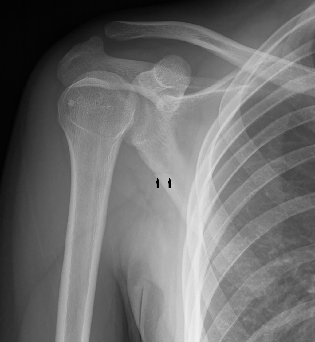
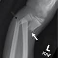
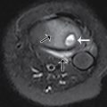
The patient had plain radiography performed. There is a transverse lucency along the lateral border of the body of the scapula, confirmed on multiple views, consistent with a nondisplaced fracture ( Figs. 54.1–54.3 ).


Stay updated, free articles. Join our Telegram channel

Full access? Get Clinical Tree


