Adverse events and complications related to interventional procedures can still occur despite the best efforts of the phlebologists. The culture of safety in health care has been extensively promoted and patient safety regulations and guidelines by specialty societies have been implemented to increase safety and improve quality. Hospitals have made substantial investments in patient safety and quality assurance in recognition of a number of complications related to surgical care. Surgical safety checklists were designed to incorporate safety protocols, minimize information loss, and promote efficient communication in the operating room. These same principles apply to phlebology procedures performed in the hospital setting or in the outpatient office.
The number of phlebology interventions performed, as well as the complexity and variety of these procedures, has increased significantly in the recent years. Despite the fact that hospitals have the infrastructure and expertise to address complications and emergencies, the office setting has become appealing to phlebologists and patients due to its convenience and cost effectiveness. Compared to hospitals, the office setting can be more pleasant to patients, usually offers improved access in convenient locations, and has lower operational costs. On the other hand, hospitals can offer prompt emergency support, electronic access to medical records and imaging, and availability of different medical specialties, which are lacking in the office setting.
In order to explore all the advantages of the office setting, and at the same time provide safe outpatient procedures, phlebology clinics should implement safety practices similar to the inpatient setting. The culture of safety in the office setting should include all aspects of patient care, from the initial contact with the patient to the assessment of posttreatment satisfaction.
A detailed medical history should be obtained and a physical examination with ultrasound evaluation of the lower extremity venous system should be performed. Pertinent information must be recorded and documented, which may include digital photographs of visible veins in the legs. The office setting should offer adequate space for staff circulation in the procedure room and light sources for the physical and ultrasound examinations. All members of the team involved with patient care should know the availability and location of emergency equipment, the protocols for cardiopulmonary emergencies, the protocols for emergency transfer of patients, and the fire evacuation protocol. For easy consult, written emergency protocols may be displayed in the procedure room.
Immediate preprocedure measures should include the following:
Review of the patient’s medical history and essential imaging studies.
Written informed consent explaining the risks, benefits, and alternatives to the procedure.
Confirmation of the patient identity and the procedure to be performed.
Adequate identification of site and side of the procedure.
Confirmation that postprocedure materials are available for immediate use, such as compression dressing or stocking.
Before discharging the patient, the following measures are recommended:
Assessment for pain and immediate postprocedure complications, such as bleeding and deep venous thrombosis (DVT).
Adequate cleaning and dressing of the treated area.
Patient education with detailed postprocedure instructions—written instructions are preferable and can be offered with contact phone numbers.
Plan for postdischarge follow-up.
9.2 Risk Assessment and Prevention in the Phlebology Clinic
9.2.1 Awareness and Understanding of Deep Venous Thrombosis
DVT poses a challenge to physicians and is a major health problem. Every physician involved with phlebology is well aware of the risks and potential consequences of DVT. Virchow’s triad of hypercoagulability, venous stasis, and endothelial injury highlight the pathophysiologic causes of DVT. Patients presenting for venous interventions often have many conditions and habits that increase their risk for the development of DVT, which are usually the same factors that led to the need for venous intervention.
Risk factors for DVT, although well known, merit repeating and should be investigated in every patient during their preoperative visit and physical examination. The risk factors include the following: history of a previous DVT; recent surgery or trauma; immobilization; malignancy; coagulopathies, either acquired or inherited—including factor V Leiden mutation; prothrombin gene mutation; antithrombin III deficiency; protein C and protein S deficiencies; antiphospholipid antibody syndrome; hyperhomocysteinemia; and plasma hyperviscosity states. The risk of DVT is also increased in pregnancy, in the postpartum state, and in patients on hormone therapy. To a lesser extent, the risk of DVT is elevated in patients with congestive heart failure, inflammatory bowel disease, obesity, and advanced age. Primary valvular insufficiency was also described as a minor risk factor for DVT. Although the risk of DVT is smaller in patients with these comorbidities than the aforementioned risk factors, the risk during venous interventions may be additive or perhaps exponentially increased due to the risks inherent to the procedure. 1, 2
Patients should be carefully screened if there is a family history of DVT or known family history of a thrombophilia. Not only are the pathologic causes of thrombosis often present in patients presenting with venous insufficiency, but also these pathologic factors are altered during each venous intervention:
Sclerosant agents entering the vein likely cause a transient hypercoagulable state.
A significant number of patients requiring venous interventions are sedentary. Pain following the procedure may further contribute to decreased activity, which leads to venous stasis.
Interventional procedures cause endothelial injury and occlusion of the superficial vein; however, untargeted endothelial injury or thrombus extension may lead to DVT.
The combination of preexisting risk factors and risk factors associated with the procedure compound the risk of DVT. 1, 2
9.2.2 Can Venous Intervention Be Performed in Patients with a History of DVT?
Venous interventions can be performed and are frequently requested for patients with a history of DVT; however, the risk of recurrence should be investigated. Stratification of the risk of recurrence can be assessed as follows:
“Provoked” DVT: It includes patients whose DVT occurred due to temporary or reversible risk factors including long-distance flight, surgery, or major trauma. The risk of recurrence is considered low.
“Unprovoked”/idiopathic DVT: It includes patients with known malignancies or coagulation disorders, or patients in whom no etiology is identified. The risk of recurrence is considered high.
The presence of residual thrombus after anticoagulation therapy also increases the risk of recurrence. Accordingly, patients who may have been initially classified in the “provoked”/low-risk group but presented with residual thrombus on a 6-month follow-up ultrasound are considered high risk. Furthermore, the risk of recurrence is higher in patients with proximal (iliofemoral) DVT in comparison with distal (femoropopliteal) DVT. 3, 4
9.2.3 To Treat or Not to Treat: Deciding to Treat or to Postpone Therapy
The myriad of DVT risk factors among patients and the variations in the severity (and the patients perception of the severity) of the varicose vein disease render the development of official guidelines a difficult and challenging task, which is why there is very little guidance in the current literature.
The decision to treat or defer therapy should take in consideration the risks and benefits of the intervention. The severity of patients’ symptoms and potential improvement following venous intervention should be carefully assessed on an individual basis, with attention to risk factors and the risk of recurrent DVT in particular.
Most physicians would consider deferring therapy if the patient has had recent major surgery, major trauma, immobilization, or pregnancy. Other conditions in which avoidance of venous interventions should be considered include patients with cancer, thrombophilia, and idiopathic DVT, unless significant improvement in the quality of life would result.
9.2.4 Preventive Measures in Patients with High Risk of DVT in Whom Venous Therapy Is Deemed Appropriate
Steps to prevent DVT. It is well known that early ambulation and mechanical or pharmacologic prophylaxis prevent DVT. The importance and necessity of early ambulation should be emphasized at length regardless of the anticipated risk of DVT. Patients should receive compression therapy, most commonly compression stocking in the postoperative period, and be educated about the importance of early and continued ambulation as well as continued compliance with compression therapy.
Awareness of DVT. Patients should be educated about the common symptoms of DVT both prior to scheduling the procedure and on the day of the procedure. This may also be available in the written postprocedure instructions provided to patients and include contact information to reach the clinical staff if symptoms occur.
Pharmacologic prophylaxis in high-risk patients. To date, there are no convincing data to support the routine use of anticoagulation prophylaxis for venous interventions such as endovenous thermal ablation or sclerotherapy. In theory, patients with hypercoagulable states may benefit from periprocedural anticoagulation; however, there is no consensus protocol for drug of choice, timing, or duration of anticoagulation. 5 Although not commonly done, some phlebologists recommend a short course of 3 to 5 days of subcutaneous administration of low-molecular-weight heparin (LMWH) in high-risk patients such as patients with cancer, thrombophilia, or idiopathic DVT. 2
Technique of venous therapy. Diligence and sound technique in theory should prevent DVT for many reasons. During endovenous thermal ablation, the tip of the delivery catheter should always be visualized to prevent activation in an undesired location causing untargeted endothelial injury. Some authors propose the use of liquid sclerosants rather than foam for high-risk patients given that foam produces greater endothelial injury and an increased transient iatrogenic hypercoagulable state. Extensive interventions performed in one setting may increase significantly the length of the procedure and recovery time. Two or more separate procedure sessions should be considered for high-risk cases to limit the time the patient is sedentary. It is a well-established practice to encourage ambulation after venous interventions because long periods of immobility could increase the risk of DVT. 2
9.3 Complications of Phlebology Interventions
Venous thromboembolic complications can result from different phlebology interventions and possibly represent the major concern during the postprocedure follow-up.
9.3.1 Superficial Thrombophlebitis
Superficial thrombophlebitis (ST) is a common inflammatory process associated with thrombosis of a superficial vein. Clinical signs and symptoms include localized pain, erythema, pruritus, edema, tenderness, and hardening of the surrounding tissue. Although there is some evidence to suggest an association between ST and venous thromboembolism, 6 further investigation is needed to clarify the correlation between ST and DVT.
In the literature, controversy exists over the definition of ST after sclerotherapy. Sclerotherapy of varicose veins induces a desired inflammatory reaction that will eventually evolve into fibrosis. Therefore, inflammation including erythema, pain, tenderness, edema, and hardening of the vein at the site of venous injection should not be interpreted as phlebitis. Consensus defines ST as superficial venous thrombosis in a noninjected vein. However, the difference between ST and an excessive but expected sclerotherapy reaction could be difficult to distinguish.
ST occurs after sclerotherapy and its incidence in the literature ranges from 0 to 46% with a mean of 5% 7, 8, 9, 10, 11; however, the real frequency of ST as defined above is unknown.
There is no difference in the rate of ST for foam compared to liquid sclerosants. However, the relative risk of ST is increased for foam sclerotherapy when compared with laser therapy and surgery. 8
Prevention
Early ambulation and the use of graduated compression stockings are thought to be important to decrease the development of ST in dependent tributary veins, although this is not based on data.
Anecdotally, ST is more commonly associated with large-diameter-dependent varicose veins or varicose tributaries following occlusion of their inflow and outflow. Therefore, concurrent phlebectomy of such veins can be performed at the time of endovenous therapy to prevent ST. 5
Treatment
Treatment aims to improve and relieve local painful symptoms but also to prevent additional thromboembolic events, which could complicate the natural course of ST. ST has been managed in most series with nonsteroidal anti-inflammatory drugs (NSAIDs), graduated compression stockings, and ambulation. However, one large placebo-controlled randomized, controlled trial of 3,002 patients showed a significant reduction in symptomatic superficial venous thrombosis in patients treated with prophylactic fondaparinux for 6 weeks. 12
The evidence on oral treatments, topical treatment, or surgery is too limited to inform clinical practice. 13 Further research is needed on the novel oral anticoagulants (NOACs) including direct thrombin and activated factor X inhibitors, LMWH, and NSAIDs to evaluate the effect on superficial venous thrombosis and ST progression, and to determine optimal doses and durations of therapy or combination therapies.
9.3.2 Deep Venous Thrombosis
DVT of the lower extremities following phlebology interventional procedures is considered a major complication since it can be associated with substantial morbidity. The development of pulmonary embolism can be life threatening. The late manifestation of postthrombotic syndrome (PTS) is also being recognized as the end-stage sequela of inadequately treated DVT. Permanently painful and swollen legs, pigmentation, skin deterioration, and venous ulceration in severe cases characterize this syndrome. In the absence of effective therapy, PTS patients have a reduced quality of life. 14
Few studies are available to evaluate the real frequency of DVT occurring after liquid sclerotherapy given that most published studies on liquid sclerosant are old and predate routine duplex ultrasound (DUS). The incidence of DVT is reportedly low in studies utilizing foam sclerosant, ranging from less than 1% to 1.8%. 8, 15, 16, 17, 18 The occurrence of DVT after endovenous thermal ablation of a superficial vein is uncommon, with reported incidences of 0.3% for laser ablation and 0.4% after radiofrequency ablation. 5
Endovenous Heat-Induced Thrombosis
Endovenous heat-induced thrombosis (EHIT) is defined as extension of thrombus from the thermal-ablated superficial vein into the adjacent deep vein. It can be illustrated as propagation of thrombus from the great saphenous vein into the common femoral vein through the saphenofemoral junction or from the small saphenous vein into the popliteal vein through the saphenopopliteal junction ( ▶ Fig. 9.1). Classification of EHIT is based on the extent of thrombus propagation into the deep venous system as follows 19, 20, 21:
Class 1: Propagation of thrombus to the level of the junction between the superficial and deep venous systems.
Class 2: Propagation of thrombus from the ablated superficial vein into the deep venous system with less than 50% intraluminal occlusion.
Class 3: Propagation of thrombus from the ablated superficial vein into the deep venous system with more than 50% intraluminal occlusion.
Class 4: Propagation of thrombus from the ablated superficial vein into the deep venous system with complete intraluminal occlusion.
Symptomatic and asymptomatic DVTs are not often distinguished in the literature, although there is a likely clinical significant difference in the outcome. Severe thromboembolic events such as proximal DVT, pulmonary embolism, or paradoxical embolization are very rare complications of phlebology interventions. Most cases of DVT diagnosed by DUS on routine follow-up are distal and asymptomatic. 7, 16
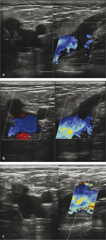
Fig. 9.1 A 55-year-old man with symptomatic varicose veins related to significant venous reflux in the left great saphenous vein (GSV). Medications in use include aspirin and Plavix indicated by the past medical history of coronary artery disease with stent placement. Endovenous laser ablation was performed without interruption of anticoagulation therapy. Immediate postprocedure ultrasound examination showed no evidence of DVT at the left common femoral vein (CFV). (a) One-week follow-up: axial and sagittal views of the left CFV at the level of the saphenofemoral junction (SFJ) showing propagation of thrombus with less than 50% occlusion—EHIT Class 2. (b) Twelve-day follow-up: axial and sagittal views of the left CFV at the level of the SFJ showing thrombus at the level of the SFJ—EHIT Class 1. (c) One-month follow-up: axial and sagittal views of the left CFV at the level of the SFJ showing thrombus in the proximal left GSV without involvement of the SFJ.
Stay updated, free articles. Join our Telegram channel

Full access? Get Clinical Tree


