Sarcomas

Chapter 32 Sarcomas


Soft tissue sarcomas


Soft tissue sarcomas are malignant tumours arising from supporting connective tissues anywhere in the body. These structures (fibrous tissue, fat, blood vessels, smooth and skeletal muscle, tendons and cartilage) are derived embryologically from the mesoderm. Sarcomas of bone also occur and are discussed in this chapter under bone tumours. Tumours of peripheral nerves are generally included. Despite arising from a wide variety of tissues, they have many similarities in pathology, clinical findings and behavior.


Pathology


Soft tissue sarcomas are rare, representing 0.4% of all cancers and 0.3% of cancer deaths. The incidence is about 0.6 per 100   000 population per year. However, they constitute 6% of tumours in children under the age of 15. Most occur in the 40–70 age group. The sex ratio is virtually the same.


Their aetiology is largely unknown. In a minority of cases, genetic factors are involved. There is, for example, an increased incidence of the tumour in association with certain genetically transmitted diseases, e.g. Gardner’s syndrome, tuberous sclerosis, Von Recklinghausen’s disease and Li–Fraumeni syndrome. Rarely, soft tissue sarcomas may occur in the children of mothers with breast cancer of early onset.


Some sarcomas are radiation induced and occur within previously irradiated areas (especially for benign angiomas). The incidence of second sarcomas among young people who have been treated with both chemotherapy and radiotherapy for Ewing’s sarcoma may reach 18%.


A large number of sarcomas have been found to have consistent chromosome abnormalities. The first demonstration of a specific gene abnormality associated with malignant transformation in man was the loss of the retinoblastoma (RB gene), a tumour supressor gene. There are alterations to the RB gene in up to 70% of soft tissue sarcomas. Chromosome translocations are the most common abnormality and are seen in virtually all cases of Ewing’s sarcoma (primitive neuroectodermal tumours). 11:22 translocation is the most common, occurring in 85% of cases. In more than 90% of synovial sarcomas, there is a chromosome 18 translocation. Such cytogenetic abnormalities may be useful diagnostically and may be of prognostic significance. It is noteworthy that translocations tend to occur most commonly in high-grade tumours.


Rhabdomyosarcoma and soft tissue Ewing’s sarcoma, although traditionally included in some lists of histological subtypes of soft tissue sarcomas, are quite different from the rest of the group, both in natural history and in being in general more chemo- and radiosensitive. Thus, the principles of treatment outlined below do not apply to these two histological types.


The World Health Organization (WHO) classification is the most commonly used, but this is extremely complex and a simplified version listing malignant tumours by their tissue of origin is presented in Table 32.1. From the point of view of management and prognosis, the histological subtype of management is generally less important than the histological grade and the size of the tumour. Grading may depend in part on the tumour subtype, but more often is assessed by scoring the degree of necrosis and mitotic index. The system described by Trojani is in regular use both in the UK and internationally. Low-grade tumours have a recognizable pattern of differentiation, relatively few mitoses, and no necrosis. High-grade tumours include all examples of certain subtypes (e.g. alveolar soft part), tumours with little or no apparent differentiation, and those with necrosis and a high mitotic rate. The tumour size is also important, and those over 5   cm are much more likely to recur locally or give rise to distant metastases.


Table 32.1 Simplified version of the WHO classification of sarcomas


Initial spread from the primary site is into adjacent tissue and along tissue planes between structures such as muscle bundles. Soft tissue sarcomas do not possess a true capsule but are often surrounded by a pseudocapsule of compressed surrounding tissues. This apparent encapsulation may tempt the surgeon to try to ‘shell out’ the tumour. Local recurrence from residual tumour at the periphery is then very likely (up to 90% within 2 years). Lymph node spread is infrequent, while blood-borne spread is common, giving rise to distant metastases, predominantly in the lungs.


Diagnosis and staging


Important features of local examination for limb or truncal sarcomas is the position of the lesion superficial or deep to the fascia at the primary tumour site and any involvement of bone, vascular or neural invasion. Regional nodes, although uncommonly involved, should be palpated.


A biopsy is required. In 90% of cases, adequate histology can be achieved by needle core biopsy. Where an incisional biopsy is required, care should be taken to make the incision longitudinal so that any subsequent radical surgery or postoperative radiation fields can include it. This is particularly important where preservation of limb function is a major consideration. Appropriate siting of the biopsy is vital if the probability of local control is to be maximized. The biopsy should ideally be carried out by the surgeon who will undertake the definitive resection. There is otherwise the risk of tumour spillage, particularly after unplanned attempts to remove deep-seated lesions. If computed tomography (CT)-guided biopsy is needed, for example for impalpable deep-seated lesions, there is also potential for needle track contamination. Close liaison with the surgeon and clinical oncologist is needed to plan the best route of biopsy. Specialist pathology review is essential in view of the complexity of these tumours and the need for careful assessment of the tumour margins.


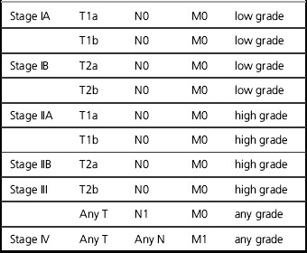
A full blood count, liver function tests, chest radiograph, plain radiographs of the tumour-bearing region (Figure 32.1) and liver ultrasound are the initial investigations. CT and magnetic resonance imaging (MRI) (Figures evolve 32.2–32.4image) scanning are important in defining the local extent and operability of the tumour. MRI is the imaging modality of choice for limb sarcomas since it provides good contrast between the tumour and adjoining normal tissues with particular regard to the tumour’s relationship with the neurovascular bundle. It also provides better multiplanar versatility in planning surgery and radiotherapy. CT can be helpful in exclusion of cortical erosion of bone. CT and MRI are also useful in confirming any subsequent local recurrence, but are not currently used for routine follow up. CT of the thorax is advisable if chest radiography is normal and radical therapy is contemplated, since it may demonstrate small-volume lung metastases (Figure evolve 32.5image). The staging system commonly adopted includes grade, size, and presence/absence of metastases (Tables 32.2 & 32.3).
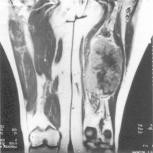


Table 32.3 Staging system for soft tissue sarcomas (UICC 2009)


T1 ≤ 5   cms
T1a Superficial
T1b Deep
T2 >5   cms
T2a Superficial
T2b Deep
N0 No lymph node spread
N1 Involvement of regional lymph nodes
M0 No distant metastases
M1 Distant metastases


Management


A multidisciplinary approach (MDT) is required since surgery, radiotherapy and chemotherapy may all have a role to play, depending on the stage, site, grade and size of the tumour. A dedicated sarcoma MDT allows treatment to be carefully coordinated and minimizes the number of surgical procedures necessary. Correct procedures for biopsy, imaging and exclusion of metastatic disease are essential. Historically, amputation was the main treatment for limb sarcomas but, in the 1970s, it was appreciated that radiotherapy and less radical surgery could provide comparable results. Limb-sparing surgery with postoperative radiotherapy achieved the same survival as amputation, albeit with a slightly higher local recurrence rate. In the past, sarcomas were regarded as relatively radioresistant tumours. It is now known that they are relatively radiosensitive, comparable to breast cancer.


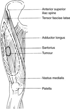
Local control while maximizing the probability of conserving limb function and long-term survival is of vital importance. For extremity limb sarcomas, 90% local control at 5 years should be the standard. The management of soft tissue sarcomas is controversial. In part, this reflects the limited number of randomized trials in this heterogeneous group of tumours. The relative rarity of non-extremity sarcomas makes the design of and recruitment to large randomized trials difficult. Few would dispute the primary role of radical surgery. If the surgical resection margins are clear, the 5-year local control for limb sarcomas is of the order of 90%. If the margins are involved, this falls to 60–80%. Involved margins are an independent risk factor for local recurrence. The extent of the resection will depend upon the site of the tumour. The more distal the tumour in the limb, the more difficult a complete excision becomes. Radical excision surgery of retroperitoneal sarcomas is also rarely feasible. The primary tumour should be resected with one anatomical plane clear of the tumour at all stages. The aim of the surgery is to achieve a wide local excision (margin ≥2   cm where possible) (Figure 32.6). The resection should include all the skin and subcutaneous tissue near to the tumour, any previous excision or biopsy scars and areas containing blood clot from previous biopsies. The tumour itself should never be actively contacted during resection. Metallic clips at the margins of the excision are helpful in planning postoperative irradiation.


Amputation for extremity sarcomas is only indicated in the following situations:


Adjuvant therapy


For low-grade tumours less than 5   cm in size which have been widely excised with clear surgical margins, no further therapy is necessary in most cases. The same applies following an amputation. However, postoperative radiotherapy is indicated in the following circumstances to reduce the probability of local recurrence (see above):


Postoperative radiotherapy conventionally is with external beam (photons or electrons), although postoperative brachytherapy with iridium-192 (42–45   Gy over 4–6 days) had a high 5-year local control rate of 90% in a randomized trial compared to 65% for high- but not low-grade lesions. There are no randomized trials comparing brachytherapy with external beam irradiation. Brachytherapy should only be carried out in specialist units undertaking a substantial number of procedures. The reason for the ineffectiveness of brachytherapy in low-grade tumours is not clear. One possibility is that the long cell cycle in low-grade tumours may result in cells not entering the radiosensitive phase of the cell cycle during the period of brachytherapy. Retroperitoneal sarcomas require special mention. They are often huge, causing dramatic distortion of the patient’s contour but rarely present as an emergency. Such cases should therefore always be discussed in the sarcoma MDT. The tumours are often dedifferentiated liposarcomas and as such have a very characteristic CT scan appearance. In these circumstances, preoperative biopsy may be eschewed, but if there is any radiological doubt, biopsy is mandated.


Surgery may require removal of many organs, spleen, one kidney, tail or body of pancreas or segments of large and/or small bowel. The placement of surgical clips around narrowly excised tumour areas can guide the clinical oncologist. The dose of post-operative radiotherapy is necessarily limited and >50   Gy is unlikely to be achievable. Recently, interest has been rekindled in the use of pre-operative RT, with very large fields being applied to a dose of 50   Gy in 1.8–2.0 Gy fractions. This has been made possible with the use of IMRT to spare radiosensitive organs such as kidney and spinal cord. An EORTC trial is recruiting now randomising to surgery alone or preoperative radiotherapy followed by radical resection 4–6 weeks later.


Protons are being used in clinical trials. Protons have a similar relative biological effectiveness (RBE) to photons. The advantage of protons is a reduction of dose to critical structures, such as the spinal cord or underlying bone. This can be done by varying the proton energy or by the use of wax compensators to alter the size of the Bragg peak and consequently the volume of tissue which is subsequently irradiated.


Not all sites are suitable for radical radiotherapy. Pelvic, thoracic or abdominal sarcomas present particular difficulties because of the morbidity that would be caused by wide-field irradiation, dose-limiting critical structures (e.g. the spinal cord). Placement of spacers in the abdominal cavity may be used to displace small bowel out of the high dose radiation field (Figures evolve 32.7 and 32.8image).


Mar 7, 2016 | Posted by in GENERAL RADIOLOGY | Comments Off on Sarcomas

Full access? Get Clinical Tree

Get Clinical Tree app for offline access