Fig. 1.1
Patient with well-differentiated neuroendocrine tumors: Octreoscan® SPECT-CT showed liver metastases
Despite greater sensitivity, limitations of Octreoscan® scintigraphy should be noted. The methodology clearly influenced the sensitivity of the examination. Routine use of planar images and SPECT-CT images of the abdomen (24 h after injection) rather than whole body images are recommended. Octreoscan®x cannot provide information on the size of the tumor. The density and type of the somatostatin receptors vary with the histologic type of the tumors: Insulinomas have a low affinity for octreotide, related to a low expression of sst subtype-2 [5]. Garin et al. [13] reported that negative Octreoscan® scintigraphy in well-differentiated endocrine tumors is negative prognostic factor.
Specificity of Octreoscan® should be noted. Some other tumoral and nontumoral diseases can show positivity of Octreoscan® [7].
Somatostatin Receptor PET-CT
Positron emission tomography (PET) scan is becoming more widely used and may be a useful localizing modality for neuroendocrine tumors as different radiolabeled substances can be used as metabolic substrate. After the development of a PET tracer for somatostatin analogs, 68Ga-DOTA-NOC (tetra-azacyclododecane tetra-acetic acid-[1-Nal3]-octreotide) has been introduced. This compound for PET imaging has a high affinity for sst2 and sst5 and has been used for the detection of NETs in preliminary studies. The uptake of 68Ga-DOTA-NOC is based on a receptor mechanism and although this has not yet been adequately assessed, it seems to have higher sensitivity for NETs than Octreoscan®, thereby increasing diagnostic accuracy. Additionally, it has several advantages over Octreoscan®: increased spatial resolution and the possibility of images with a short uptake time (60 min), and relatively easy synthesis [14].
The two other compounds most often used in functional imaging with PET are 68Ga-DOTATOC and 68Ga-DOTATATE. 68Ga-DOTATOC and 68Ga-DOTATATE possess similar diagnostic accuracy for detection of NET lesions. The increasing availability of 68Ga somatostatin analogs PET-CT now offers superior accuracy for localization and functional characterization of NETs. However, studies are needed to enable imaging of NET with optimal targeting of tumor receptors.
18F-DOPA PET-CT
Fluorodihydroxyphenylalanine-(18F-FDOPA) PET is a recent imaging modality used to localize neuroendocrine tumors [15]. These tumors have the ability to produce biogenic amines and polypeptide hormones, and they take up and decarboxylate their amine precursors, L-dihydroxyphenylalanine. 18FDOPA-PET appears to be a major tool for the management of carcinoid tumors with excellent diagnostic performances (65–96 %) related to these capacities to concentrate amino acids inside the vesicules of cytoplasmatic space through metabolic mechanism. 18FDOPA-PET is less sensitive and less useful for the management of noncarcinoid tumors (Fig. 1.2).
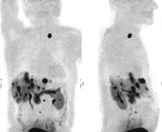

Fig. 1.2
Patient with well-differentiated neuroendocrine tumors: 18FDOPA PET-CT showed multiple liver metastases and sus-clavicular left lymph node
18FDG PET-CT
Although 18F-2-Deoxy-d-glucose (18F-FDG) PET is the most widely used and accepted type of PET in clinical oncology, it has limited use in well-differentiated tumors such as NETs due to their low expression of glucose transporters and low proliferative activity. However, several studies have evaluated 18F-FDG PET-CT in well-differentiated NET [13, 16, 17]. Garin et al. reported that 18FDG uptake is a poor prognostic factor in NETS, in relation to tumor aggressiveness and is related to a lower overall survival (Fig. 1.3).
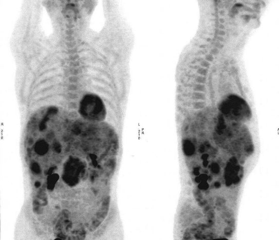

Fig. 1.3
Patient with well-differentiated neuroendocrine tumors grade 2 (KI 67: 15 %).18FDG-PET-CT showed liver metastases and mesenteric lymph node
Conclusions
All of these performances highlight the significant contribution of the scintigraphic procedures from a diagnostic point of view and the management of therapy of patients with NETs. PET imaging could be of major interest for the diagnosis, evaluation of progression and treatment response in NETs. 18FDG-PET even though still not validated, carries major prognostic information and may influence determination of the optimal therapeutic strategy. The role of 18F-DOPA is clearly recommended before surgery for the detection of carcinoid tumors. The different new somatostatin analogs with 68Ga radiolabeling must be evaluated.
Stay updated, free articles. Join our Telegram channel

Full access? Get Clinical Tree


