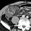
Thin (usually < 3 mm) pseudocapsule encasing inflamed mesentery (tumoral pseudocapsule sign)
Cluster of mildly enlarged mesenteric nodes
Usually located in left upper quadrant mesentery
Mesenteric vessels and nodes have halo of spared fat (fat ring or fat halo sign)
•
Chronic phase
Chronic fibrosis results in discrete fibrotic soft tissue mass with desmoplastic reaction
“Stellate” appearance with calcification within mass
Encasement of mesenteric vessels (especially portomesenteric veins) with resultant collaterals
–
Small bowel wall edema and mucosal hyperemia as result of lymphatic/venous obstruction and ischemia
Tethering of bowel loops can lead to bowel obstruction
•
Retroperitoneal and mesenteric lymphoma
•
Desmoid tumor (fibromatosis)
•
Carcinomatosis (mesenteric metastases)
•
Primary visceral malignancy
•
Inflammatory pseudotumor
•
Unknown etiology, but associations with prior surgery, trauma, infection, autoimmune diseases, and malignancies
•
Classified histologically into 3 types or stages based on predominant tissue type in mass
Mesenteric lipodystrophy: Fat necrosis > inflammation or fibrosis
Mesenteric panniculitis: Acute inflammation ± fat necrosis > fibrosis
Retractile mesenteritis: Fibrosis/retraction > inflammation or fat necrosis
•
Acute mesenteritis can be cause of acute abdominal pain
•
Natural history or frequency of progression to chronic fibrotic phase not well understood
Most patients have stable or slowly progressing disease without symptoms
•
Common complications in chronic phase include bowel obstruction, urinary tract obstruction, and bowel ischemia
•
Treatment: Immunosuppressive therapy should be attempted initially
Limited role for surgery, as surgical excision is very difficult in advanced cases due to vascular involvement
•
Retractile mesenteritis, fibrosing mesenteritis, mesenteric panniculitis, mesenteric lipodystrophy, liposclerotic mesenteritis, systemic nodular panniculitis, xanthogranulomatous mesenteritis
•
Idiopathic inflammatory and fibrotic disorder affecting mesentery of unknown etiology
•
Best diagnostic clue
“Misty mesentery” with surrounding pseudocapsule, clustered prominent mesenteric lymph nodes, and halo of spared fat surrounding nodes and vessels
•
Location
Most common site: Root of jejunal mesentery
–
90% involve small bowel mesentery and primarily to left of midline (jejunal mesentery)
Occasionally: Colon (transverse or rectosigmoid)
Rarely: Peripancreatic, omentum, and retroperitoneum
•
Morphology
Mostly characterized by mixture of mesenteric inflammation, fat necrosis, and fibrosis
•
Key concepts
Uncommon, benign, inflammatory process involving mesenteric fat
Classified histologically into 3 types or stages based on predominant tissue type in mass
–
Mesenteric panniculitis: Acute inflammation ± fat necrosis > fibrosis
–
Mesenteric lipodystrophy: Fat necrosis > inflammation or fibrosis
–
Retractile mesenteritis: Fibrosis/retraction > inflammation or fat necrosis
Retractile mesenteritis
–
Considered as final, chronic form, with collagen deposition, fat necrosis, fibrosis, and tissue retraction
Often associated with other idiopathic inflammatory disorders (> 1 condition may be present)
May coexist with malignancy (e.g., lymphoma, breast, lung, colon cancer, and melanoma)
•
Acute mesenteritis (mesenteric panniculitis and lipodystrophy)
Often associated with misty mesentery: Increased attenuation of mesentery with fat stranding and induration
–
Nonspecific finding that can be seen with other etiologies
–
Usually located in left upper quadrant mesentery
–
Often discrete fat-attenuation “mass” with increasing soft tissue component as disease progresses
Thin (usually < 3 mm) pseudocapsule encasing inflamed portion of mesentery (tumoral pseudocapsule sign)
Cluster of mesenteric nodes (only rarely enlarged > 1 cm) within misty mesentery
–
Mesenteric vessels and nodes have halo of surrounding spared fat (fat ring or fat halo sign)
•
Chronic phase (retractile mesenteritis)
Chronic fibrosis results in discrete fibrotic soft tissue mass with desmoplastic reaction
–
Usually located in root of mesentery (left upper quadrant)
–
Mass often has “stellate” appearance with internal calcification (and within adjacent lymph nodes)
Can rarely show internal cystic or necrotic components
–
Encasement of mesenteric and collateral vessels with frequent narrowing/occlusion of portomesenteric veins
Collaterals or engorged vessels may be present
Small bowel wall edema and mucosal hyperemia as result of lymphatic/venous obstruction and ischemia
–
Tethering of bowel loops can lead to bowel obstruction
•
Variable signal intensity due to varying elements of inflammation, fat necrosis, calcification, and fibrosis
•
Acute mesenteritis (panniculitis and lipodystrophy)
T1WI: Mixed signal intensity
T2WI: Mixed signal intensity (usually mildly hyperintense)
•
Chronic phase (retractile mesenteritis): Imaging findings of mature fibrotic reaction
T1WI: Decreased signal intensity
T2WI: Very low signal intensity
Get Clinical Tree app for offline access
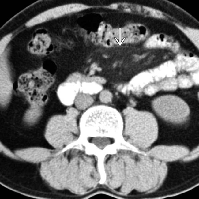
 .
.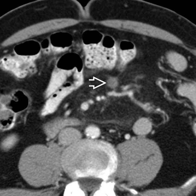
 . No diagnosis was made at this time, and the patient was not given treatment.
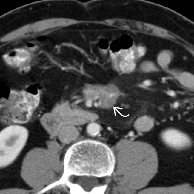
 in the mesentery that encases and narrows the mesenteric vessels.
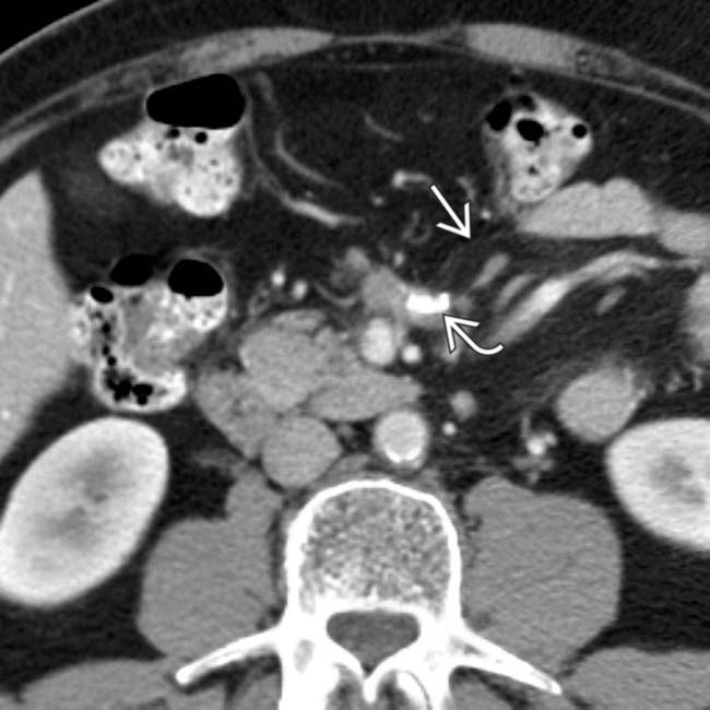
 within the fibrotic mesenteric mass. The infiltrated mesentery and pseudocapsule
  are still evident. This case illustrates progression of the disease over time.
 Often associated with misty mesentery: Increased attenuation of mesentery with fat stranding and induration
 Thin (usually < 3 mm) pseudocapsule encasing inflamed portion of mesentery (tumoral pseudocapsule sign)
 Often associated with misty mesentery: Increased attenuation of mesentery with fat stranding and induration
 Thin (usually < 3 mm) pseudocapsule encasing inflamed portion of mesentery (tumoral pseudocapsule sign)
 Narrowing/occlusion of flow in mesenteric vessels


 Narrowing/occlusion of flow in mesenteric vessels


