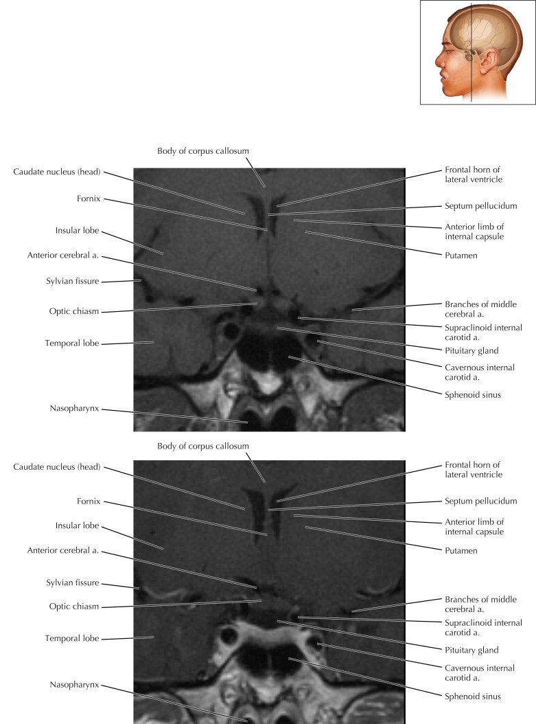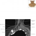

Sella Turcica Coronal 2
Diagnostic Considerations
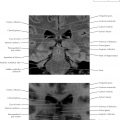
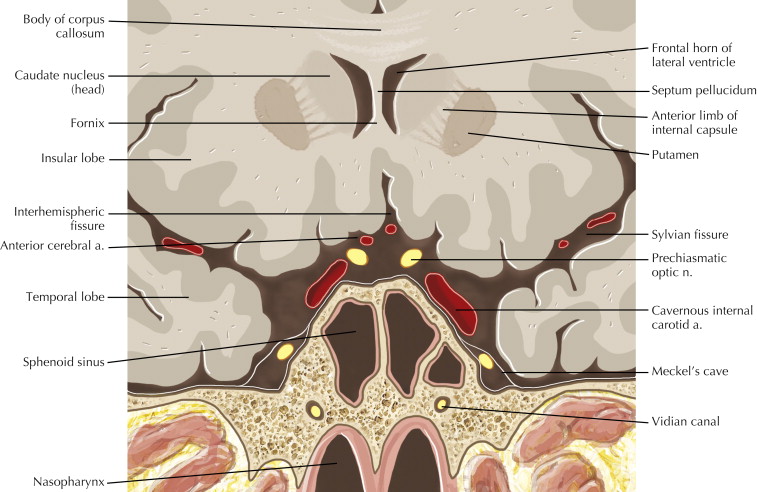
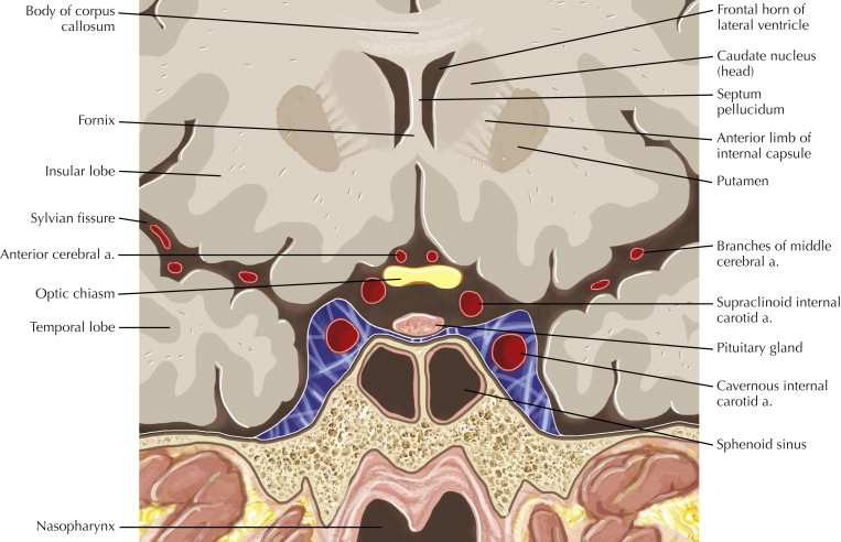
The best magnetic resonance sequence to identify a pituitary lesion is coronal, thin, 3-mm magnified postcontrast images of the sella turcica. The sella is a transverse depression crossing the midline at the superior surface of the sphenoid bone that contains the pituitary gland, or hypophysis. Pituitary adenomas are typically less enhancing than the normal gland. Precontrast imaging is used to determine that the apparent enhancement is not intrinsic high T1-weighted signal. Intrinsically high T1 signal is most frequently seen with fat, some forms of protein, acute hemorrhage (methemoglobin), and microcalcifications.
Sella Turcica Coronal 2