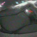
Fig. 15.1
(a) Near-infrared camera system (PDE, photodynamic eye). (b) PDE system. (c) The camera unit was covered with a sterilized transmissive cover. (d) The camera was handled directly by the surgeon
Before performing laparotomy, a fine needle (26 gauge) was inserted into the submucosal layer via the anus, and ICG dye (Diagnogreen, Daiichi Sankyo Co., Ltd., Tokyo, Japan) was gently injected at four sites along the dentate line circumferentially. The total amount of injected dye was 1 ml (5 mg, 0.5 % ICG) in each patient. After the rectal cancer was resected, the lateral vesical and obturator spaces were opened via the extraperitoneal approach between the lateral aspect of the internal iliac vessels and the pelvic wall. The adipose tissue surrounding the common-internal iliac arteries and obturator space was observed with PDE, which allowed for the identification of SN in the lateral pelvic region.
15.2.3 Division of the Lateral Pelvic Region
According to the Japanese Classification of Colorectal Carcinoma [19], the lateral pelvic region can be classified into three divisions: (1) the common iliac region along the common iliac artery, (2) the internal iliac region along the internal iliac artery, and (3) the obturator region along the obturator nerve, artery, and vein (Fig. 15.2).
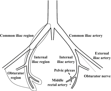
Fig. 15.2
Diagram showing the division of the lateral pelvic region
15.2.4 Statistical Analysis
The statistical analyses were performed using the StatView version 5.0 software package (Abacus Concepts, Berkeley, CA). The data are presented as the mean ± standard deviation and were analyzed using the χ 2 test for discrete variables. If the sample size was small, Fisher’s exact test was used. A P value of <0.05 denoted the presence of a statistically significant difference.
15.3 Results
15.3.1 Patient Characteristics
The patients included 36 males (67.9 %) and 17 females (32.1 %), with an average age of 59.8 ± 11.5 (range, 32–83) years. The anal edge of all tumors at the lower level was located either at or below the peritoneal reflection. The distance from the dentate line to the lower border of the tumor was 2.9 ± 2.1 (range, −2.0 to 6.0) cm, and the tumor size was 4.6 ± 1.7 (range, 1.2–11.0) cm. Most tumors were moderately differentiated adenocarcinomas (N = 38), followed by well-differentiated adenocarcinomas (N = 13) and poorly differentiated adenocarcinomas (N = 2) (Table 15.1).
Table 15.1
Clinicopathological findings of the 53 patients with rectal cancer
Age (years) | 59.8 ± 11.5 (32–83) |
Gender | |
Male | 36 (67.9 %) |
Female | 17 (32.1 %) |
Distance from dentate line (cm) | 2.9 ± 2.1 (−2.0–6.0) |
Tumor size (cm) | 4.6 ± 1.7 (1.2–11.0) |
Histological grade | |
Well | 13 (24.5 %) |
Mod | 38 (71.7 %) |
Por | 2 (3.8 %) |
Primary tumor | |
pT1 | 3 (5.7 %) |
pT2 | 13 (24.5 %) |
pT3/T4 | 37 (69.8 %) |
Regional lymph nodes | |
pN0 | 25 (47.2 %) |
pN1 | 17 (32.1 %) |
pN2 | 11 (20.8 %) |
pTNM stage | |
I | 10 (18.9 %) |
II | 15 (28.3 %) |
III | 22 (41.5 %) |
IV | 6 (11.3 %) |
Lymphatic invasion | |
No | 20 (37.8 %) |
Yes | 33 (62.3 %) |
Venous invasion | |
No | 14 (26.4 %) |
Yes | 39 (73.6 %) |
15.3.2 Feasibility
No adverse events related to the ICG injections were noted. As previously described [20], the optimal concentration of ICG was 0.5 %, and a total of 1 ml (5 mg) of ICG solution was injected. PDE clearly visualized the LNs and lymphatic vessels that received ICG; however, the PDE-positive LNs were not detected with the naked eye. The LNs and lymphatic vessels not seen under ordinary white light were visualized on PDE in the surrounding adipose tissue around the common-internal iliac arteries and obturator space (Fig. 15.2). The preparation of the device, injection of the ICG solution, and observation of the fluorescence images took less than 30 min.
15.3.3 Detection of Lateral SNs
The LNs and lymphatic vessels that received ICG appeared as shining fluorescent spots and streams on the fluorescence images of PDE. The lateral SNs were successfully identified in 49 (92.5 %) of the 53 patients; the total number of lateral SNs was 94, and the number of lateral SNs per patient was 1.9 ± 0.8 (range, 1–4). Of the 94 SNs, 51 (54.3 %) were detected in the internal iliac region, 40 (42.5 %) were detected in the obturator region, two (2.1 %) were detected in the external iliac region, and one (0.1 %) was detected in the common iliac region. The parameters age, sex, distance from the dentate line, tumor size, histological grade, depth of the primary tumor, upward LN status, pTNM stage, lymphatic invasion, and venous invasion did not affect the successful identification of the lateral SNs (Fig. 15.3, Table 15.2).
 ICG Fluorescent Image-Guided Surgery in Head and Neck Cancer
 Regional Lymph Node Dissection Assisted by Indocyanine Green Fluorescence Lymphography and Angiography for Stage III Melanoma
 Indocyanine Green Fluorescence-Navigated Sentinel Node Navigation Surgery (SNNS) for Cutaneous Malignant Melanoma and Extramammary Paget’s Disease
 Anatomical Hepatectomy Using Indocyanine Green Fluorescent Imaging and Needle-Guiding Technique
 Basic Aspects of ICG Fluorescence Imaging of the Liver
 Principle and Development of ICG Method


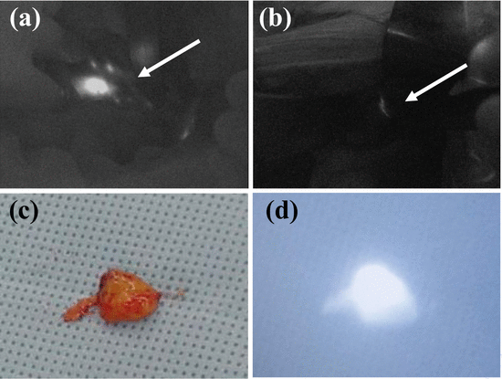
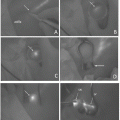
Fig. 15.3
Detection of SNs around the left internal iliac artery. (a) On PDE, the LNs that received ICG appeared as shining fluorescent spots (arrow). (b) On PDE, the lymphatic vessels that received ICG appeared as shining fluorescent streams (arrow). (c) The SNs that received ICG were not stained green in the naked eye examination. (d) The same SNs that received ICG appeared as shining fluorescent spots on PDE
Table 15.2
Clinicopathological features and rate of success in identifying lateral SNs
Clinicopathological features | No. of cases examined (N = 53) | No. of SNs detected in cases (N = 49) | Success rate (%) | P value |
|---|---|---|---|---|
Age | ||||
<60 years old | 30 | 28 | 93.3 | >0.9999 |
≥60 years old | 23 | 21 | 91.3 | |
Gender | ||||
Male | 33 | 31 | 93.9 | 0.6274 |
Female | 20 | 18 | 90.0 | |
Distance from the dentate line | ||||
<3.0 cm | 22 | 20 | 90.9 | 0.1241 |
≥3.0 cm | 31 | 29 | 89.0 | |
Tumor size | ||||
<4.5 cm | 24 | 22 | 91.6 | >0.9999 |
≥4.5 cm | 29 | 27 | 93.1 | |
Histological grade | ||||
Well/Mod | 51 | 47 | 92.2 | >0.9999 |
Por | 2 | 2 | 100 | |
Depth of the primary tumor | ||||
pT1 | 3 | 3 | 100 | 0.8772 |
pT2 | 13 | 12 | 92.3 | |
pT3/T4 | 37 | 34 | 91.9 | |
Upward LN status | ||||
negative | 28 | 26 | 92.9 | >0.9999 |
positive | 25 | 23 | 92.0 | |
pTNM stage | ||||
I | 10 | 9 | 90 | 0.7766 |
II | 15 | 14 | 93.3 | |
III | 22 | 21 | 95.5 | |
IV | 6 | 5 | 100 | |
Lymphatic invasion | ||||
No | 20 | 19 | 95.0 | >0.9999 |
Yes | 33 | 30 | 90.9 | |
Venous invasion | ||||
No | 14 | 13 | 92.9 | >0.9999 |
Yes | 39 | 36 | 92.3 | |
Well well-differentiated adenocarcinoma, Mod moderately differentiated adenocarcinoma, Por poorly differentiated adenocarcinoma, LN lymph node, SN sentinel node
Related posts:
ICG Fluorescent Image-Guided Surgery in Head and Neck Cancer
Regional Lymph Node Dissection Assisted by Indocyanine Green Fluorescence Lymphography and Angiography for Stage III Melanoma
Indocyanine Green Fluorescence-Navigated Sentinel Node Navigation Surgery (SNNS) for Cutaneous Malignant Melanoma and Extramammary Paget’s Disease
Anatomical Hepatectomy Using Indocyanine Green Fluorescent Imaging and Needle-Guiding Technique
Basic Aspects of ICG Fluorescence Imaging of the Liver
Principle and Development of ICG Method
Stay updated, free articles. Join our Telegram channel
Full access? Get Clinical Tree
