• Treatment: Sigmoidoscopic decompression of obstruction ± stabilization via rectal tube insertion
Usually followed by surgical resection of sigmoid colon
DIAGNOSTIC CHECKLIST
• Rule out other causes of distal colonic obstruction
• Dilated sigmoid colon in inverted “U” shape with absent haustra; “beaking,” whirl sign, northern exposure sign
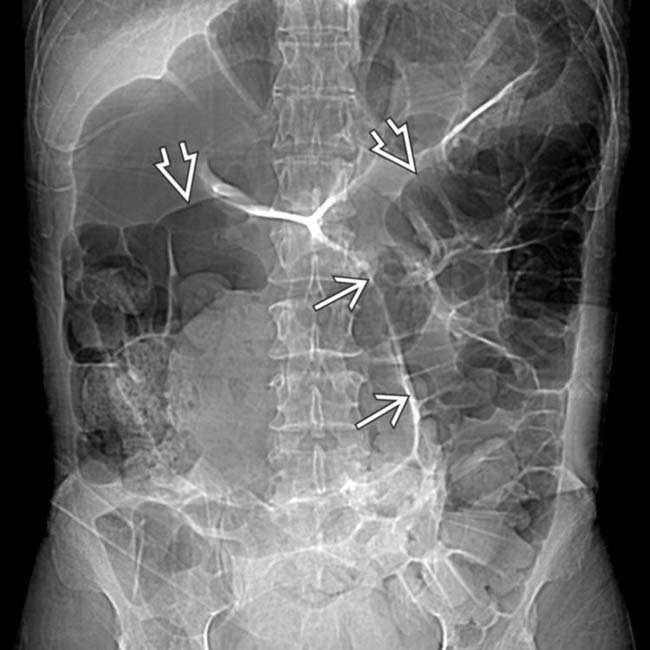
(Left) Supine film of the abdomen shows marked dilation of the sigmoid colon. The sigmoid is folded back upon itself, and the apposed walls of the redundant sigmoid colon form the “seam” of the football (or coffee bean) shape. The sigmoid extends into the upper abdomen above the transverse colon .
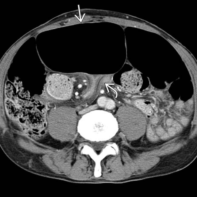
(Right) Axial CECT in the same case shows the dilated sigmoid lumen with abrupt narrowing at its base .
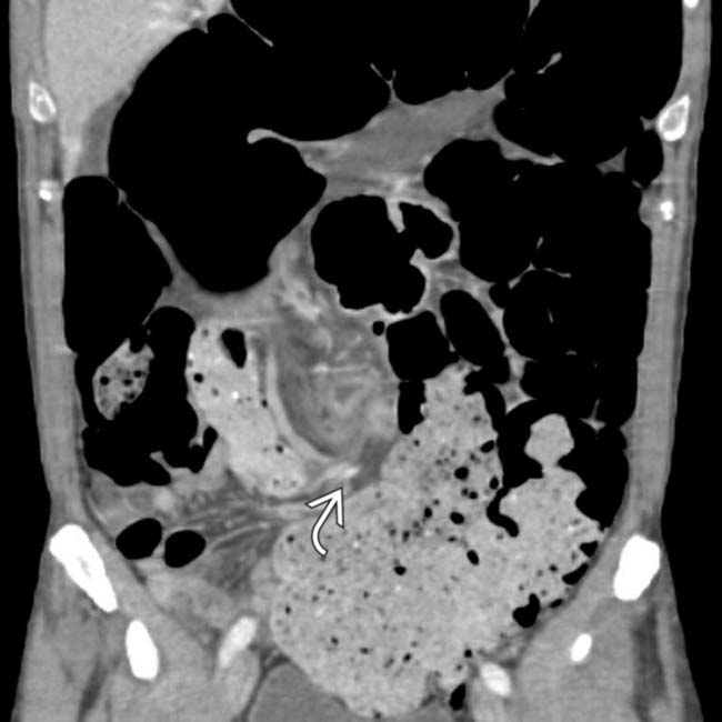
(Left) Coronal reformatted CT in the same patient shows twisting and displacement of the base of the sigmoid colon and its mesentery . The dilated colonic segments upstream from the volvulus may be easier to distinguish on coronal sections.
(Right) Another CT section in this case shows the whirl sign of twisted colon and vessels at the base of the sigmoid mesentery.
TERMINOLOGY
Definitions
• Torsion or twisting of sigmoid colon around its mesenteric axis
IMAGING
General Features
• Best diagnostic clue
Dilated sigmoid colon with inverted “U” configuration and absent haustra
• Location
Midline; directed toward RUQ or LUQ; elevation of hemidiaphragm
Radiographic Findings
• Radiography
Sigmoid volvulus
– Diagnostic in 75% of cases
– Vertical dense white line: Apposed inner walls of sigmoid colon pointing toward pelvis
– Closed loop obstruction: Segment of bowel obstructed at 2 points
– Gas in proximal small intestine and colon; absence of gas in rectum
– Absent rectal gas in spite of prone or decubitus views


 form the “seam” of the football (or coffee bean) shape. The sigmoid extends into the upper abdomen above the transverse colon
  .
.
 with abrupt narrowing at its base
  .
.
 . The dilated colonic segments upstream from the volvulus may be easier to distinguish on coronal sections.
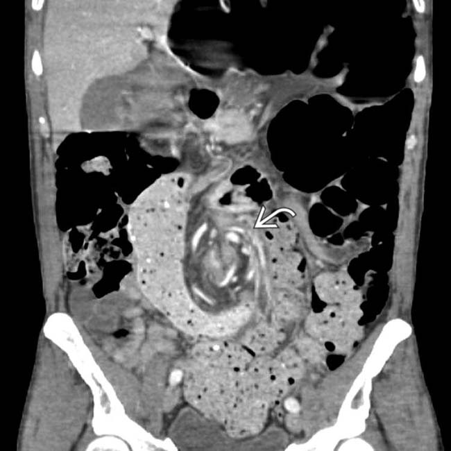
 of twisted colon and vessels at the base of the sigmoid mesentery.


