Localization: It usually arises in the metaphyses of the long bones, beneath the growth plate, but along with patient growth, the cyst migrates to the diaphysis. The most common location is the proximal humerus, followed by proximal femur.
Clinical: Per se asymptomatic, the simple bone cyst becomes painful due to frequent pathologic fractures. Sometimes it presents as an incidental finding.
Imaging: The characteristic radiographic appearance is that one of a pure osteolysis, thinning, and “inflating” the cortex. Bone septa, usually present, give the appearance of a multiloculated cyst. When fracture occurs, a small fragment of the wall may be seen on X-rays in the cavity, also known as “fallen leaf” sign.
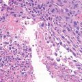
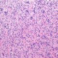
Pathology: Grossly it appears as a cavity filled by serous fluid similar to synovial fluid. When the cyst is growing, a thin membrane made of flattened connective cells with few osteoclasts lines the wall. When the lesion is inactive, cuboidal cells line the cavity, while cholesterol slits, hemosiderin deposits, scattered giant cells, and osteoblasts are present along the wall. About 20 % of the cyst walls contain clumps of pink, granular amorphous material that resembles cementum or fibrinoid material that is quite diagnostic of SBC when seen.
Course and Staging: Bone cysts are staged according to their activity. Lesions abutting to the growth plate usually have an internal pressure >30 cm H2O, a shiny membrane, and overall clinical features of activity, being considered stage 2. Lesions located far from the physis usually have an internal lower pressure, a thicker membrane, and overall features of inactivity, being considered stage 1. Occasionally the fracture may induce spontaneous healing of the cyst.
Treatment and Prognosis: Percutaneous methylprednisolone injections appear to have a reasonable success rate in the majority of active cysts. Effectiveness of injection in latent lesions is debatable and case-by-case approach suggested. Curettage and bone grafting +/− internal fixation in fracture or risk of fracture.
Key Points
Clinical | Incidental findings or pain if fractured
Related posts:Stay updated, free articles. Join our Telegram channel
Full access? Get Clinical Tree
 Get Clinical Tree app for offline access

|