Case presentation
A 16-year-old female presents to the emergency department with complaints of right wrist and hand pain following a fall on an outstretched hand during sports practice after school. Vital signs are age appropriate and her physical examination is significant for tenderness in the anatomic snuffbox and the radial portion of the wrist. Grip strength and range of motion in the wrist are both reduced in the injured hand. She is neurovascularly intact in the right upper extremity and has no other significant findings on physical examination.
Imaging considerations
Imaging is often obtained in acute traumatic orthopedic injuries. Plain radiographs are often the first-line imaging modality, although additional modalities including computed tomography (CT) and magnetic resonance imaging (MRI) may be required.
Plain radiography
In the case of a possible scaphoid fracture, a standard wrist series should be obtained. Plain radiographs should be the initial imaging modality; however, scaphoid fractures may go undetected on initial plain radiography, with false-negative reads on the initial imaging reported as high as 20%–54%. Scaphoid fractures may not be visualized on a standard three-view wrist series but are best seen on specialized scaphoid views; one of these is an anteroposterior scaphoid view taken with the wrist in full pronation and with 30 degrees of ulnar deviation, but other scaphoid views include posteroanterior views, also with ulnar deviation.
Magnetic resonance imaging
MRI has greater sensitivity and specificity for scaphoid fracture than CT does and MRI shows greater accuracy than other modalites do. , MRI can detect bone marrow edema within hours of the injury. MRI is also able to accurately detect injuries in the other surrounding osseous and soft tissues.
Computed tomography
CT is sensitive and highly specific for the diagnosis of scaphoid fractures. This imaging technique can be used immediately following the trauma; however, CT is not as sensitive as MRI or bone scan for scaphoid fracture and cannot as conclusively rule out a scaphoid fracture. CT can be useful in patients who have contraindications to receiving an MRI.
Bone scan (bone scintigraphy)
While skeletal scintigraphy is an additional option for identifying a scaphoid fracture, it must be performed at least 72 hours postinjury. It is also important to consider that while bone scan sensitivity for scaphoid fracture is the highest at 99%, it only has an 86% specificity. Bone scan also requires the injection of radioactive material and has the highest radiation exposure of the imaging options, which is a significant consideration, especially in the pediatric population. Bone scans have the benefit of being able to detect other bony injuries.
Case conclusion
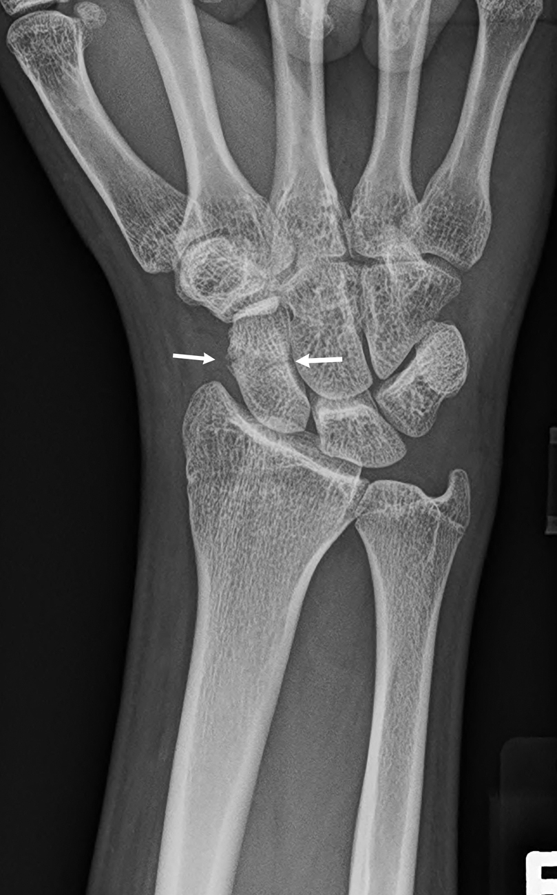
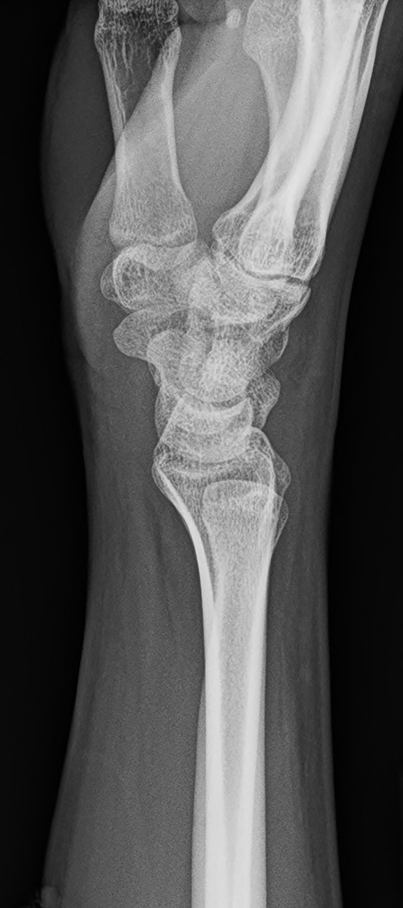
Plain radiographs of the wrist were obtained in the emergency department. These images demonstrate a nondisplaced transverse fracture of the scaphoid ( Figs. 60.1–60.3 ). The other visualized bones are normal.