Fig. 22.1
(a–d) B-cell non-Hodgkin’s lymphoma in a 55-year-old man: (a) axial spin-echo T1-weighted MR image; (b) axial spin-echo T1-weighted MR image with fat suppression; (c) axial spin-echo T1-weighted MR image after gadolinium contrast injection with fat suppression; (d) sagittal spin-echo T2-weighted image. The T1-weighted image shows a diffuse infiltrating mass with a slightly higher signal intensity than adjacent muscle (a). With fat suppression the lesion becomes more hyperintense, and the extent of the lesion in the infraspinatus, trapezoid, and deltoid muscle becomes apparent (b). After intravenous contrast administration, there is a diffuse moderately heterogeneous contrast enhancement providing a good differentiation between muscle fibers and tumor on the T1-weighted image (c). On the T2-weighted image, the lesion is better depicted owing to the hyperintense appearance relative to the low signal intensity of muscle, illustrating the extent of tumoral involvement with a sharp transition zone to the surrounding trapezoid muscle (d). The T2-weighted image and the T1-FS-weighted image after gadolinium confirm an intact aspect of the cortical bone of the scapula with a sharp demarcation between the relative hyperintense aspect of the tumor and the cortical bone of the scapula (c, d)

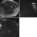
Fig. 22.2
(a–e) Giant B-cell lymphoma in a 68-year-old woman: (a) axial CT image after IV contrast injection; (b) axial spin-echo T1-weighted MR image; (c) axial spin-echo T1-weighted MR image after gadolinium contrast injection with fat suppression; (d) coronal spin-echo T1-weighted MR image after gadolinium contrast injection with fat suppression; (e) axial spin-echo STIR-weighted image. CT image after contrast shows a butterfly-shaped lesion consisting of a soft tissue mass enveloping the cortical bone of the iliac crest. The lesion shows a homogeneous contrast enhancement making the lesion mildly hyperdense in comparison to the gluteal and iliac muscles (a). The T1-weighted image shows no apparent destruction of the iliac wing. The lesion is rather homogeneous presenting with a signal intensity similar to the surrounding muscle structures (b). Both T1- and T2-weighted images with fat suppression illustrate focal areas of abnormal bone marrow due to permeative bone infiltration characteristic of lymphoma without apparent bone destruction. The presentation is typical for the “wraparound” sign seen in bone and soft tissue lymphoma (c–e). The hyperintensity of the lesion after fat suppression allows better evaluation of the tumor extent and better delineates the transition of tumoral tissue to normal gluteal and iliac muscle fibers. The tumor enhances homogeneously showing minor internal septation (c, d)

Fig. 22.3
(a–d) Biopsy-proven non-Hodgkin’s lymphoma of the quadriceps muscle in a 64-year-old woman: (a) axial spin-echo Tl-weighted MR image; (b) axial spin-echo T2-weighted MR image; (c) coronal; (d) axial spin-echo Tl-weighted MR images after gadolinium contrast injection. On a Tl-weighted image, the lesion presents as a nodular mass, infiltrating the fascia and superficial fibers of the rectus femoris, vastus intermedius, and adductor longus muscles. The lesion has a slightly higher signal intensity than adjacent muscle. There is an extensive tumoral spread into the subcutis and cutis resulting in venous obstruction and superficial varices (a). After contrast injection there is a diffuse, moderately heterogeneous contrast enhancement providing a good differentiation between muscle fibers and tumor on Tl-weighted images (c, d). On a T2-weighted image, the extent of the lesion can be depicted better owing to the hyperintense appearance relative to the low signal intensity of muscle (b). The T2-weighted or fat-suppressed images also allow better appreciation of venous involvement

Fig. 22.4
(a–d) A 66-year-old man with left sciatica presenting with a type B non-Hodgkin’s lymphoma in and around the sciatic nerve: (a, b) ultrasound; (c) axial spin-echo Tl-weighted MR image; (d) sagittal spin-echo T2-weighted MR image. A nodular type of lesion is found on ultrasound between the hamstring muscles. The lesion is sharply demarcated and presents with similar but more heterogeneous echogenicity to that of the hamstring muscles (a). In the caudal border of the lesion, hyper-echogenicity is due to infiltration of the tumor in the surrounding fat (b). On T1-weighted images, there is a nodular lesion in the center of the hamstrings along the neurovascular bundle. The lesion has a mildly hyperintense to isointense appearance, making differentiation with adjacent muscle groups relatively difficult (c). On T2-weighted images, a mildly hyperintense, bead-like lesion extending along the nerve root with irregular fat planes toward the biceps femoris (caput longum) muscle is seen

Fig. 22.5
(a–e) A 64-year-old man with a palpable soft tissue mass at the left upper arm (non-Hodgkin’s lymphoma): (a) radiography of the left humerus; (b) CT scan after iodinated contrast injection; (c, d) sagittal spin-echo T1- and T2-weighted MR images; (e) axial spin-echo T1-weighted MR image after gadolinium contrast administration. The initial radiographic examination reveals a permeated aspect of the cortex of the left humerus of a lengthy area with linear and irregular periosteal abnormalities (a). Contrast-enhanced CT scan shows a diffuse iso-attenuating infiltrating mass, whereas the borders of the lesion cannot be distinguished from the adjacent muscle fibers. The density of the bone marrow is abnormal (b). The higher signal intensity of the tumor on corresponding T1- and T2-weighted images (c, d) is indicative of a diffuse infiltrating mass extending along the humeral shaft and infiltrating the brachioradial and triceps compartments. The bony “wraparound” sign of the tumor resulting in periosteal reaction at the humeral diaphysis and in bone marrow involvement is considered highly suggestive of soft tissue lymphoma. T1-weighted images after gadolinium contrast administration allow evaluation of tumor extension into different compartments and exclusion of invasion of the neurovascular bundle (e)
Two different morphological patterns are encountered: a more lobular, which is usually more superficial, and a deeper, diffuse infiltrative pattern. In both cases lesions behaved aggressively, often infiltrating multiple muscle groups or compartments, with no respect for compartment boundaries. Multicompartmental involvement is noted in 50 % of cases often extending into the subcutaneous tissues in about 40 % of cases [2, 35] (Fig. 22.6a–e). The transition of the tumor is irregular without cleavage planes toward adjacent muscle fibers (Figs. 22.3 and 22.4). Infiltration along the neurovascular bundle (Fig. 22.2) and extension through subcutaneous strands are often present (five out of eleven cases in our series). These strands are hypointense on TI-WI and mildly hyperintense on T2-WI. There is little or no gadolinium-DTPA enhancement in these strands corresponding to perilesional edema. Often there is an encasement of major vascular structures. MRI is the imaging technique of choice for demonstrating neurovascular encasement and defining the extent of cortical bone and marrow involvement, found in 7 out of 24 cases in the series of Suresh et al. [31]. Lymphoma can infiltrate and/or surround the cortical bone with changes in the signal intensity of the adjacent marrow. In these cases, differentiation between secondary soft tissue lymphoma by extracortical spread of primary bone lymphoma and primary soft tissue lymphoma with secondary bone involvement is often not possible (Fig. 22.5). The first possibility is supported by findings of Hicks and the second by findings of Moulopoulos [14, 24].

Fig. 22.6
Subcutaneous B-cell lymphoma at the right upper arm, MRI (a–e) and PET (f) (Reprinted with permission from reference Vanhoenacker FM, Baten A, Vandeputte V (2009) Imaging findings of a cutaneous B-Cell Lymphoma. JBR-BTR 92:285–288). (a) Axial T1-WI demonstrating a well-demarcated slight hyperintense mass involving the subcutaneous fat and the skin at the anterior-lateral aspect of the upper arm. The lesion has an intimate relationship with the fascia of the biceps muscle. There is a small additional subcutaneous lesion within the subcutaneous tissue of the chest wall with similar SI characteristics (arrow). (b) Coronal fat-suppressed T2-WI of the right upper arm shows a large fusiform mass (white arrows), which is of intermediate signal intensity. (c) Axial fat-suppressed T1-WI. The lesion has a slightly higher signal intensity than the muscle. The small additional subcutaneous lesion within the subcutaneous tissue of the chest wall is seen as well (arrow). (d) Axial fat-suppressed T1-WI after intravenous injection of gadolinium contrast. Marked and homogeneous enhancement of the lesion as well of a subcutaneous nodule at the back of the trunk is demonstrated (arrow). (e) Axial subtraction image of the enhanced and nonenhanced MR images at a slightly different level. The homogeneous enhancement pattern is more obvious. Notice also the presence of two enhancing satellite nodules at the subcutaneous tissue of the chest wall (arrows). (f) [18F]fluorodeoxyglucose (FDG) positron emission tomography. Notice a focal area of intense FDG uptake at the right upper arm (white arrow), as well as two additional foci of increased uptake at the right chest wall (black arrowhead) and a fourth small subcutaneous lesion at the right thigh (black arrow)
Moulopoulos [24] reviewed a series of 13 stage 4 lymphomas with bone involvement. If tumor was present on either side of the bony cortex but the contour of the affected bone was preserved, it was described as “wrapped around” (Fig. 22.3). This wraparound sign is indicative of a primary soft tissue lymphoma and is not found in patients with myeloma or metastatic disease.
Hicks et al. [14] showed that the absence of cortical destruction in the case of a soft tissue mass adjacent to a bone lymphoma does not exclude the possibility of an extension of the primary bone disease into adjacent soft tissues. In their study of primary bone lymphoma, they showed the presence of intracortical channels which are filled with tumor.
In those studies, however, the intraosseous component preceded the development of a soft tissue mass or was very extensive in proportion to the soft tissue mass itself. In our series the soft tissue component of the tumors was accompanied by only a mild endomedullary component in four cases (Fig. 22.3). The soft tissue component was obviously more extensive than the bone marrow abnormalities. Intracortical channels were not shown either before or after intravenous gadolinium-DTPA administration. Consequently these lesions are considered primary soft tissue lymphoma with secondary bony involvement.
22.5.2 Nuclear Medicine and Hybrid Imaging
Similar to systemic lymphoma [18F]fluorodeoxyglucose positron emission tomography (FDG-PET) shows a marked accumulation of radionuclide, and therefore FDG-PET or FDG-PET-CT is strongly recommended before treatment for patients with routinely FDG-avid, potentially curable lymphomas to better delineate the extent of disease; however, currently it is not routinely indicated because of limitations imposed by cost and restriction in availability [5] (Fig. 22.6f).
A diffuse, widespread accumulation of radioisotope in the affected muscle can also be seen in rhabdomyolysis [20]. Rhabdomyolysis can be a sequela of lymphoma, as a result of the tendency of malignant lymphoma to increase the amount of muscle damage. Muscle damage may be caused by diffuse infiltration into muscles, by involvement of multiple neighboring muscle compartments and metastasizing into other soft tissues, as well as a sequela of cytotoxic actions. This cytotoxic action with acute tubular necrosis can account for the number of patients reported with renal dysfunction associated with muscle lymphomas.
Related posts:


Stay updated, free articles. Join our Telegram channel

Full access? Get Clinical Tree


