Fig. 24.1
Intramuscular metastases. (a) Gray-scale and (b) color Doppler 12–5 MHz US images in patient with previously diagnosed malignancy (lung adenocarcinoma): well-defined lobulated shape hypoechoic nodules located within the muscle. Color Doppler imaging shows a hypervascular pattern. (c) Contrast-enhanced CT demonstrates an intramuscular (M. left erector spinae) lesion with central low attenuation and rim enhancement (arrow)

Fig. 24.2
Intramuscular metastasis from choroid melanoma. (a) Gray-scale US reveal a small solid homogeneously hypoechoic nodule with spiculated margins in the subcutaneous tissue of the abdominal wall. (b) Axial contrast-enhanced helical CT image shows enhancing lesion in right external and internal oblique muscles (arrow) and a left psoas extension of a vertebral metastasis
Sonographic guidance provides a safe, rapid, and accurate method for localizing superficial soft tissue masses suggestive of metastatic disease and then guiding needle biopsies to acquire cytologic material for a definitive diagnosis [1].
23.5.3 CT
CT is not an ideal method for characterization of soft tissue metastases. Furthermore, soft tissue metastases cannot be reliably distinguished from the primary soft tissue sarcoma [51].
CT appearance of soft tissue metastases is widely variable: “abscess-like” lesion with rim enhancement and central hypo-attenuation, focal masses with homogeneous contrast enhancement, diffuse metastatic infiltration manifested as soft tissue swelling and diffuse heterogeneous contrast enhancement, multifocal calcification, and intramuscular bleeding with muscle enlargement and area of high attenuation [45]. The most common appearance of soft tissue metastases on contrast-enhanced CT is that of a rim-enhancing mass with central hypo-attenuation (Figs. 24.1, 24.2, 24.3, 24.4, and 24.5) [29].


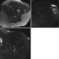
Fig. 24.3
17-year-old woman with osteosarcoma. Contrast-enhanced abdominal CT scan shows enhancing lesion in right external and internal oblique muscles with multifocal calcifications (arrows)
Fig. 24.4
Skeletal muscle metastasis in patient with caecal mucinous adenocarcinoma (asterisk). (a) Contrast-enhanced CT demonstrates intramuscular lesion with central low attenuation and rim enhancement (arrow). (b) Axial fat-suppressed (FS) T1-weighted gadolinium-enhanced image shows well enhancing mass in left psoas including central necrosis. (c) Two months later, nonenhanced CT shows enlargement of the left psoas muscle with multifocal calcifications
Fig. 24.5
Intramuscular metastases. (a) US image in patient with previously diagnosed osteosarcoma: well-defined hypoechoic mass with multifocal hyperechoic areas located within the vastus intermedius. (b) Nonenhanced and (c) contrast-enhanced CT demonstrates an intramuscular (vastus intermedius) lesion with multifocal calcifications and homogeneous contrast enhancement. (d) Contrast-enhanced CT at a lower level shows a subcutaneous round mass with homogeneous contrast enhancement
CT may guide lesion biopsy and direct appropriate local radiation.
23.5.4 MRI
MR imaging is the preferred technique for muscle and soft tissue assessment, even though MRI appearances of soft tissue metastasis are not specific [13, 52].
On MRI, soft tissue metastases are of low or intermediate signal intensity compared to normal muscle on T1-weighted sequences and high signal intensity on T2-weighted sequences [17, 22, 42].
However, high signal intensity of metastases on T1-weighted images has been also described, particularly in malignant melanoma metastases because they contain melanin and may often bleed [54]. High signal intensity has also been reported in metastases of renal cell carcinoma, but its cause is still unknown [20, 35].
After administration of contrast medium, most muscle metastases show marked heterogeneous enhancement related to tumor necrosis (Fig. 24.4). In addition, extensive peritumoral enhancement has been reported as one of the characteristic features of IM [13, 48].
Edema of surrounding soft tissue is common [6]. Erosion of the adjacent bone might rarely be observed on MRI [17].
Diffusion-weighted imaging (DWI) is considered as a useful method in the assessment of tumor cellularity in soft tissue sarcomas and can be used as a noninvasive tool to monitor treatment responses [36, 49]. In soft tissue metastases, Surov described low or moderate ADC values and moderate to high signal intensity on diffusion images [44].
23.5.5 Radionuclide Imaging
Technetium-99 m-labeled nuclear scan can show increased uptake within the soft tissue mass [48]. 18F-FDG PET/CT was demonstrated to have a higher sensitivity compared with MRI in detecting soft tissue metastases [10 31]. However, there were false positives in few cases, which should be taken into consideration. PET/CT, which performs anatomical and metabolic assessment, is very sensitive in the detection of soft tissue metastases and provides more information for clinical tumor staging. Compared to CT, PET is more sensitive because it detects soft tissue lesions before the density and morphology of the soft tissues are altered, providing a clear view of highly metabolic nodules within the soft tissue. 18F-FDG PET/CT imaging may reveal the primary tumors of the soft tissue metastases, which is helpful for differential diagnosis.
23.6 Biopsy
After appropriate imaging, early percutaneous biopsy is recommended. It is advocated to biopsy in case of solitary or late metastases and in case of unknown primary tumor. Biopsy is not recommended in case of a disseminated disease.
23.7 Differential Diagnosis
Several benign tumors and soft tissue sarcoma cannot be differentiated radiologically from metastases [5, 16, 28]. Psoas involvement by metastatic disease is frequently confused with psoas abscess or hematoma, both clinically and radiographically. Therefore, confirmation of soft tissue metastases can only be established by histopathology.
23.8 Prognosis
The presence of soft tissue metastasis influences the primary tumor treatment. Reported studies show that the survival of patients with soft tissue metastases ranges from less than 9 months to not more than 3 years after soft tissue metastases diagnosis, although survival up to 5 years has been reported in some cases [24]. The presence of a skin metastasis in a lung cancer patient indicates a poor prognosis, with a median survival of 2–4 months [10, 25, 38].
23.9 Treatment
The treatment of soft tissue metastases depends on their localization and clinical presentation as well as on the prognosis related to the primary tumor. Therapeutic options include observation, radiotherapy, chemotherapy, and surgery. For painful masses in the context of a widespread metastatic disease, radiotherapy, chemotherapy, or both may be indicated based on the primary tumor, the organ involved, the extent of involvement, symptoms attributable to the various sites, the patient’s age, and health status [6].
Conclusion
Radiologists should know various imaging and clinical findings of soft tissue metastases and suspect it in cases of painful and multiple masses. The diagnosis is based on imaging techniques, especially MRI, CT, and 18F-FDG PET/CT. However, the confirmation of diagnosis is based on the histological examination if necessary.
Key Points
Soft tissue metastases are uncommon and their prevalence varies in autopsy series from 6 to 17.5 %.
The thigh muscles, iliopsoas, and paraspinous muscles are the most frequent affected sites.
The most frequent symptoms are pain, palpable mass, swelling, and cutaneous erythema.
Radiographs and CT are not ideal methods for characterization of soft tissue metastasis.
On US, soft tissue metastases appear as well-circumscribed hypoechoic and hypervascularized masses.
On MRI, soft tissue metastases are of low or intermediate signal intensity compared to normal muscle tissue on T1-weighted sequences and high signal intensity on T2-weighted sequences.
It is advocated to biopsy solitary or late metastases and in case of unknown primary tumor.
References
1.
2.
Batson OV (1940) The function of the vertebral veins and their role in the spread of metastases. Ann Surg 112:138–149CrossrefPubMedPubMedCentral
Related posts:


Stay updated, free articles. Join our Telegram channel

Full access? Get Clinical Tree


