Localization: The most frequent localization is in the long bones: distal femur, proximal humerus, and proximal tibia. It originates from the metaphysis, but, with skeletal growth, it tends to move toward the diaphysis. In the trunk, more frequently involved are the scapula and ilium. Osteochondroma is exceptional in the hand and foot, and it does not occur in bones originating from membranous ossification (skull) nor in the epiphyses and carpal and tarsal bones (except the calcaneus).
Clinical: Swelling is the main symptom, slowly increasing during skeletal growth. Osteochondroma is usually painless. More rarely, pain may be due to bursitis or activity-related discomfort, especially in large lesions. Occasionally, a bursa forms on the osteochondroma, due to chronic friction; when fluid collection occurs in this bursa, an apparent and rapid increase of the volume, accompanied by pain, can arise the suspicion of a malignant change. Exceptionally, osteochondroma compresses a peripheral nerve or the dural sac, thus causing neurological symptoms; or it rubs against a large artery thus producing a false aneurysm (femoropopliteal artery). Also exceptionally, as a result of traumatic fracture of its stalk, osteochondroma becomes painful and mobile, clinically simulating a muscular ossification or a loose articular body.
Imaging: Osteochondroma is a bony excrescence with well-defined limits, having a thin outer cortex and an internal cancellous structure. The pathognomonic radiographic feature is that the cortex of the host bone flares into the cortex of the osteochondroma, and the cancellous bone of the osteochondroma blends with the cancellous bone of the metaphysis. In large lesions, areas or rarefied bone may alternate with irregular blotches of intense radio density, due to remnants of calcified cartilage, focal thickening of bone trabeculae, and bone necrosis. Some are pedunculated with a globose, cauliflower-like summit or with a sharp hornlike extremity. Others have a broad sessile base. Pedunculated osteochondromas are usually inclined toward the diaphysis. Rarely osteochondroma reaches huge sizes (even 15–20 cm), which are not proof of malignancy. By chronic compression, osteochondroma can cause scalloping and bowing of an adjacent bone. CT, MRI, sometimes angiography, and ultrasound may be useful (a) to confirm diagnosis in atypical cases, (b) for preoperative planning (relationship with the vascular bundle), and (c) in cases of suspect malignant change (thickness of the cartilage cap, fluid collection in reactive bursitis). Isotope scan is hot in active osteochondromas during childhood and adolescence and remains weakly positive or becomes negative after skeletal maturity, becoming again positive in malignant changes and in some bursitis.
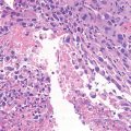
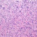
Pathology: In children, a cartilage cap covers osteochondroma with a thickness ranging from a few mm to 1 cm or more, and it appears as a light blue cartilage similar to that of the physeal plate. In the adult, this cap decreases in thickness and in some areas it disappears; residual cartilage is white and similar to articular cartilage. Limits of the cartilage with the underlying bone are well-defined. The inner part of osteochondroma is irregularly cancellous, with fatty or occasionally hemopoietic marrow. When osteochondroma is covered by a bursa, this may contain serous or hematic effusion, rarely osteocartilaginous loose bodies. In its active stage, the cartilage cap presents, although irregular, the same features of the normal growth plate. Some cellularities, plumpness of the nuclei, and hypertrophy of the cells are to be expected in children and adolescents. The bony trabeculae of osteochondroma are originated by enchondral ossification of cartilage. Cancellous bone may include remnants of calcified cartilage and/or areas of necrotic bone. Some parosteal reactive ossifications (so-called exostosis of the great toe, bizarre osteocartilaginous pseudotumor of the hand) may be composed by proliferating cartilage undergoing enchondral ossification and thus may be somewhat similar to osteochondroma; these lesions, however, lack the overall above-mentioned clinico-radiographic features characteristic of osteochondroma.
Related posts:


Stay updated, free articles. Join our Telegram channel

Full access? Get Clinical Tree


