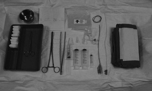
Sonohysterography

23 Sonohysterography


Chiou Li Ong

Indications


  • Abnormal uterine bleeding

    • Postmenopausal
    • Premenopausal

  • Abnormal endometrial finding on conventional ultrasonography

    • Abnormal thickening
    • In symptomatic postmenopausal women, endometrial thickness exceeding 5 mm (measurement of the two layers of endometrium)
    • Sonohysterography (SHG) can help to triage patients to “blind” dilatation and curettage, or hysteroscopy and biopsy, depending on whether diffuse or focal abnormalities of the endometrium are found.
    • Criteria for abnormal endometrial thickness in premenopausal and asymptomatic postmenopausal women are less clear.
    • Upper limits for asymptomatic postmenopausal women is around 6–8 mm, and should be correlated with hormonal or tamoxifen therapy.

  • Poor visualization of the endometrium

    • May be encountered as a result of the axial orientation of the uterus or previous cesarean section
    • SHG may be indicated in symptomatic patients.

  • Leiomyomas with distortion of the endometrium
  • Diagnostic workup prior to assisted reproductive techniques
  • To confirm presence of retained products of conception

Contraindications


Absolute Contraindications


  • Acute pelvic inflammatory disease
  • Pregnancy

Relative Contraindications


  • Hydrosalpinx
  • Bleeding
  • Patients with dilated fallopian tubes should have their examinations deferred until they have had an adequate course of antibiotics.
  • Although bleeding is not a contraindication to the examination, the presence of blood clots and fibrin strands may present problems in interpretation.

Types of Procedures


Gynecological Pelvic Ultrasound


  • Pelvic ultrasonography is the primary imaging modality for the assessment of the endometrium.
  • Two main approaches to pelvic ultrasonography: transabdominal for panoramic view and transvaginal for greater resolution and greater diagnostic accuracy
  • SHG – a useful adjunct and more specific in diagnosing endometrial pathology

Hysterosalpingography


  • X-ray technique that involves ionizing radiation and iodinated contrast medium

Hysteroscopy


  • More invasive and may be reserved for patients with focal abnormalities that are found on SHG.

Preprocedural Evaluation and Preparation


  • A brief gynecological/medical history is taken.
  • Patients with abnormal vaginal discharge suggestive of a pelvic infection should have their examination rescheduled until after the infection has been treated.
  • Prophylactic antibiotics are recommended in patients with chronic pelvic inflammation or cardiac valvular problems.
  • History of allergy to latex, if latex transducer covers or catheters are used
  • Use of a checklist is helpful.
  • Nonsteroidal antiinflammatory medication may be given prior to the procedure to minimize cramping.

Timing of Procedure


  • It should be performed during the early proliferative phase of the menstrual cycle, typically fourth to sixth day, when the endometrium is thinnest and least likely to produce false-positive results.
  • Avoid doing the procedure beyond the 10th day of the patient’s cycle
  • Patients who have irregular menstrual periods, in whom their real menses cannot be determined, may be given a course of progesterone by the gynecologist to induce “medical curettage” prior to the procedure.

Choice of Catheters


  • Nonballoon catheters (examples)

    • Infant feeding tubes (5- to 6-French) usually adequate
    • Intrauterine insemination catheter

  • Balloon catheters

    • Foley catheter
    • Commercially available hysterosalpingography catheters

  • Choice of catheter

    • Balloon catheters provide a seal on the cervical canal and help prevent rapid expulsion of fluid.
    • Study by Dessole and colleagues found no significant difference among six commonly used catheters.

Technique


Mar 10, 2016 | Posted by in ULTRASONOGRAPHY | Comments Off on Sonohysterography

Full access? Get Clinical Tree

Get Clinical Tree app for offline access