– Right heart failure, portal hypertension, sickle cell disease (in acute setting), and splenic vein thrombosis
Hematologic
– Polycythemia vera, myelofibrosis, and hemoglobinopathies
Inflammatory/infectious
– Mononucleosis and HIV/AIDS most common infections to result in splenomegaly
– Sarcoid may result in mild splenomegaly with multiple small hypodensities in liver and spleen
Space-occupying lesions
– Space-occupying masses in spleen do not commonly cause splenomegaly and are more likely to replace normal splenic tissue
– Cysts, lymphoma, metastases, and primary splenic tumors may very rarely cause splenomegaly
Storage and infiltrative disorders
– Primary or secondary hemochromatosis, amyloidosis, and glycogen storage diseases
CLINICAL ISSUES
• Complications include splenic rupture and hypersplenism
Hypersplenism: Hyperfunctioning spleen removes normal RBC, WBC, and platelets from circulation
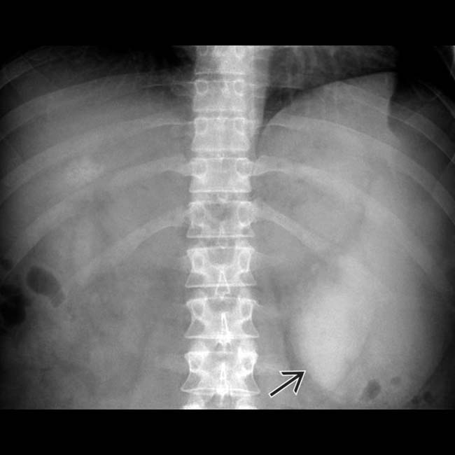
(Left) Frontal radiograph demonstrates “fullness” in the left upper quadrant. The inferior edge of an enlarged spleen is evident.
(Right) Coronal T2 MR demonstrates a markedly enlarged spleen in a patient with myelodysplastic syndrome. The most common causes of massive splenomegaly are cirrhosis/portal hypertension, lymphoma, chronic myelogenous leukemia, extramedullary hematopoiesis, myelofibrosis, and Gaucher disease.
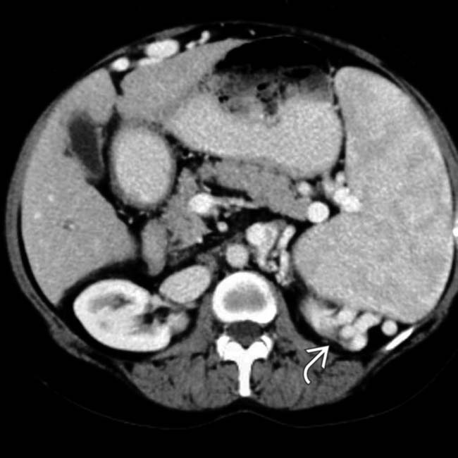
(Left) Axial CECT shows a small, cirrhotic liver with widened fissures and signs of portal hypertension, including splenomegaly and varices . In most patients with splenomegaly, there are clues as to the underlying cause on the imaging study, as in this case.
(Right) Coronal CECT in an asymptomatic patient demonstrates a mildly enlarged spleen with multiple ill-defined hypodense nodules in a patient with known sarcoidosis. Lymphoma and metastatic disease could have a very similar appearance.
TERMINOLOGY
Abbreviations
• Splenomegaly (SMG)
• Hypersplenism (HS)
Definitions
• SMG: Splenic enlargement caused by a number of different underlying congestive, hematologic, inflammatory/infectious, neoplastic, or infiltrative disorders
• Hypersplenism: Syndrome consisting of splenomegaly and pancytopenia in which bone marrow is either normal or hyperreactive
IMAGING
General Features
• Best diagnostic clue
↑ volume of spleen with convex medial border
• Size
No consensus on absolute size thresholds for SMG: Different sources suggest different measurements
Normal spleen is ≤ 13 cm in length
– Width and breadth are usually ≤ 6 and 8 cm, respectively
Splenic index: Normally 120-480 cm³ (product of length, breadth, and width of spleen)
Splenic weight: Splenic index × 0.55
– Normal weight: 100-250 g
SMG: Anteroposterior (AP) diameter > 2/3 distance of AP diameter of abdominal cavity
• Morphology
SMG is often associated with abnormal contour of spleen, including rounding of poles and convexity of medial border
Radiographic Findings
• Radiography
Normal-sized spleen usually not visualized
SMG: Splenic tip below 12th rib
Marked SMG may displace stomach medially
Displacement of splenic flexure of colon (splenic flexure usually anteromedial to spleen)
Calcification within or adjacent to spleen
CT Findings
• SMG is usually due to 1 of 5 general etiologies
• Congestive
Right heart failure: Cardiomegaly with distension of hepatic veins/IVC and passive hepatic congestion
Portal hypertension: Splenomegaly with varices, nodular shrunken liver, ascites, and other signs of portal hypertension
Splenic or portal vein occlusion or thrombosis (often due to pancreatitis or pancreatic tumors)
Sickle cell disease
– Acute phase: Diffusely decreased splenic density with splenomegaly
– Chronic phase: Development of small autoinfarcted, calcified spleen
• Hematologic
Polycythemia vera
Leukemia
Myelofibrosis: SMG due to extramedullary hematopoiesis
– May be associated with other signs of extramedullary hematopoiesis (such as paraspinal soft tissue masses)
Hemoglobinopathies: May cause splenomegaly (thalassemia) or small, infarcted spleen (sickle cell [SC])
Acute splenic infarction: Global or wedge-shaped hypoenhancement of splenic parenchyma
• Inflammatory/infectious
Mononucleosis
Hepatitis: Splenomegaly due to viremia or cirrhosis with portal hypertension
AIDS: SMG may reflect chronic viremia, opportunistic infection, or lymphoma
IV drug abuse: SMG due to chronic low-level sepsis
Tuberculosis, histoplasmosis: Multifocal low-density granulomas acutely that heal as calcified foci
Sarcoidosis: Often associated with innumerable small hypodense splenic granulomas, ± upper abdominal lymphadenopathy, ± hepatomegaly with similar hypodense hepatic granulomas
Collagen vascular or autoimmune diseases
– Rheumatoid arthritis, scleroderma, etc.
– Felty syndrome: Rheumatoid arthritis, splenomegaly, and granulocytopenia
Splenectomy may be necessary to treat hypersplenism
Only gold members can continue reading. Log In or Register to continue


 Space-occupying lesions

 of an enlarged spleen is evident.
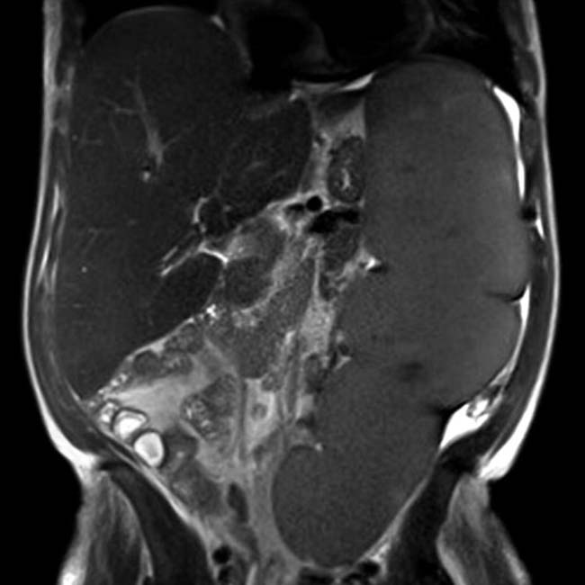
 . In most patients with splenomegaly, there are clues as to the underlying cause on the imaging study, as in this case.
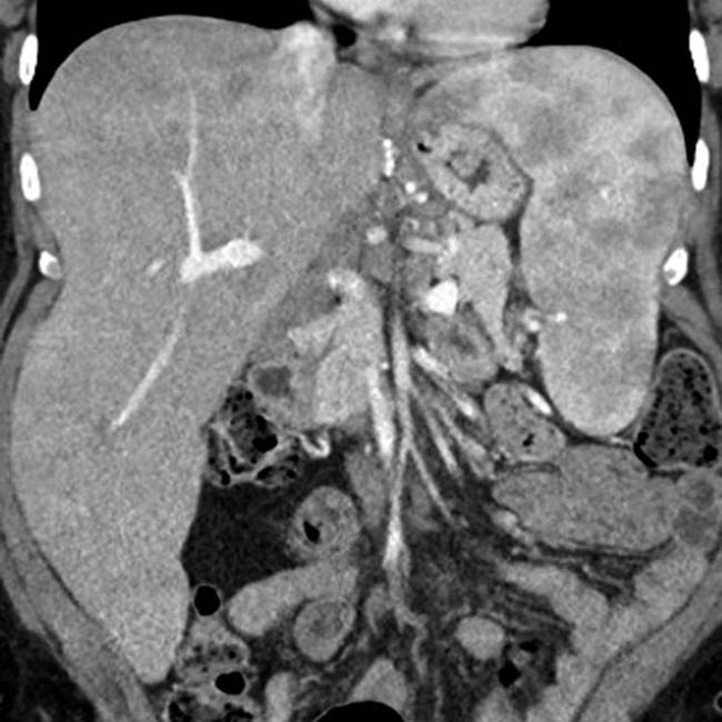


 Right heart failure: Cardiomegaly with distension of hepatic veins/IVC and passive hepatic congestion
 Portal hypertension: Splenomegaly with varices, nodular shrunken liver, ascites, and other signs of portal hypertension

 Hemoglobinopathies: May cause splenomegaly (thalassemia) or small, infarcted spleen (sickle cell [SC])


 Sarcoidosis: Often associated with innumerable small hypodense splenic granulomas, ± upper abdominal lymphadenopathy, ± hepatomegaly with similar hypodense hepatic granulomas
 Collagen vascular or autoimmune diseases


