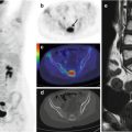
Fig. 26.1
A 12-year-old girl with spondylodiscitis was evaluated in a PET study, performed while she was on antibiotic therapy. Sagittal (a, b) and axial (c) CT and PET/CT fusion images with bone window show inhomogeneous FDG uptake corresponding to the intervertebral disk between the tenth and eleventh vertebrae. Antibiotic treatment was continued
Teaching Point
PET and MRI are of similar accuracy in the diagnosis of spondylodiscitis, supporting the use of PET when MRI findings are doubtful or the exam is not possible [9]. PET is more accurate and more specific than MRI in assessing the therapeutic response of spondylodiscitis and in some cases is preferable to MRI in the determination of when medical treatment can be safely discontinued.
References
1.
Ziegelbein J, El-Khoury GY (2011) Early spondylodiscitis presenting with single vertebral body involvement: a report of two cases. Iowa Orthop J 31:219–224PubMed
2.
Budnik I, Porte L, Arce JD, Vial S, Zamorano J (2011) Espondilodiskitis caused by Kingella kingae in children: a case report. Rev Chilena Infectol 28(4):369–373PubMedCrossRef
Stay updated, free articles. Join our Telegram channel

Full access? Get Clinical Tree


