Stage
T
N
M
G
IA
T1a
T1b
N0
N0
M0
M0
G1, GX
G1, GX
IB
T2a
T2b
N0
N0
M0
M0
G1, GX
G1, GX
IIA
T1a
T1b
N0
N0
M0
M0
G2, G3
G2, G3
IIB
T2a
T2b
N0
No
M0
M0
G2
G2
III
T2a, T2b
Any T
N0
N1
M0
M0
G3
Any G
IV
Any T
Any N
M1
Any G
The T stage is determined by maximum tumor diameter (T1/T2) and location relative to the muscle fascia (a/b). N and M stages describe the absence or presence of regional lymph node and distant metastasis, respectively. For grading (G), the former four-grade system has been replaced by a three-grade system as proposed by the French Federation of Cancer Centers (Fédération Nationale des Centres de Lutte Contre le Cancer (FNCLCC)) [7, 11, 22, 41].
The AJCC/UICC stage is predictive of overall survival. The general prognosis is better with well-differentiated tumors, smaller and superficial tumors, and in the absence of nodal involvement and distant metastasis. The presence of distant metastasis at any site indicates stage IV disease regardless of histologic tumor grade. The 5-year survival rates of extremity soft tissue sarcomas are 80–90 % at stage I and II, 56 % at stage III, and less than 20 % at stage IV [17, 22, 34, 41].
8.2.2 MSTS/Enneking Staging System
The staging system of the Musculoskeletal Tumor Society (MSTS) developed by Enneking (Table 8.2) is applicable to both soft tissue and bone sarcomas and has a more surgical orientation [14, 31, 41, 42]. It has been designed before advanced imaging techniques and adjuvant therapy were routinely used.
Stage | G | T | M |
|---|---|---|---|
IA | G1 | T1 | M0 |
IB | G1 | T2 | M0 |
IIA | G2 | T1 | M0 |
IIB | G2 | T1 | M0 |
III | G1 or G2 | T1 or T2 | M1 |
Like the AJCC/UICC system, it includes the grading (G) of the tumor but only distinguishes between low-grade and high-grade lesions. This two-tiered grading was favored, because it could better be related to surgical procedures (wide vs. radical excision) [41]. Regional lymph node metastasis is disregarded in this system. The size of the tumor is only an indirect variable, because the T stage is determined by grouping the lesions as either intracompartmental (T1) or extracompartmental (T2) rather than by absolute size [14, 41, 42,]. This makes the MSTS system best applicable to sarcomas located in the extremities [41].
8.2.3 Tumor Size
Tumor size has an influence on the prognosis of soft tissue sarcomas, mainly because it is more difficult to eradicate larger tumors surgically. The designation of 5 cm used as a cutoff value between T1 and T2 lesions in the AJCC/UICC staging system is however more or less arbitrary [22, 41]. To measure the true dimensions of a soft tissue sarcoma on MR imaging might be difficult, if the mass is surrounded by an indistinct pseudocapsule. The pseudocapsule is a peripheral zone of reactive tissue that can develop due to compression, hyperemia, and chronic inflammation of soft tissue adjacent to the sarcoma and might contain tumoral digitations and satellites [5, 6, 41]. On MR images, it is seen as a zone of peritumoral “edema” [5]. The true tumor diameter (with exclusion of the pseudocapsule) can be best measured on T2-weighted or contrast-enhanced MR images without fat suppression (Fig. 8.1).

Fig. 8.1
(a, b) T stage according to AJCC/UICC and MSTS. (a) Coronal contrast-enhanced T1-weighted SE and (b) axial T2-weighted TSE images of a patient with a pleomorphic sarcoma show a large tumor in the posterior compartment of the thigh. Tumor diameter is measured with exclusion of the pseudocapsule as demonstrated. According to AJCC/UICC, the tumor would be classified as T2b (>5 cm, deep tumor). If the MSTS system is used, the tumor represents a T1 lesion, because it is confirmed to one anatomic compartment (intracompartmental), albeit the compartmental boundaries are bulged by the mass effect
If measurements of resected specimens at pathologic examination are used as standard of reference, tumor size is often “overestimated” on preoperative MR images, especially for large masses. Possible explanations for this effect are changes in tumor shape and suspension of perfusion after resection, rupture of cystic tumor components, fragmentation, and shrinkage due to tissue fixation on pathologic preparation [4, 10, 20, 28].
8.2.4 Tumor Depth
Tumor depth is defined in the AJCC/UICC staging system on the basis of tumor location relative to the muscle (deep) fascia (Fig. 8.2). Superficial tumors are located subcutaneously and entirely superficial to the muscle fascia without invading it. Deep tumors lie either entirely beneath the muscle fascia, above the fascia and invading it, or both above and beneath the fascia [42]. On MR imaging, it is particularly difficult to differentiate close contact without fascial invasion and true invasion in tumors with subcutaneous location. Tumor depth might therefore be overestimated rather than underestimated on radiologic assessment [20].

Fig. 8.2
(a–d) T stage according to AJCC/UICC and MSTS. (a) Axial contrast-enhanced T1-weighted TSE image with fat suppression shows intramuscular leiomyosarcoma (arrow) in the anterior compartment of the thigh. AJCC/UICC: T1b, MSTS: T1. (b) Axial T2-weighted TSE image demonstrates pleomorphic sarcoma (arrow) in the posterior compartment of the thigh. AJCC/UICC: T2b, MSTS: T1. (c) Axial contrast-enhanced T1-weighted TSE image with fat suppression shows pleomorphic sarcoma (arrow) in the subcutaneous tissue involving to the muscle fascia and deep compartment. AJCC/UICC: T2b, MSTS: T2 (d) Axial T2-weighted TSE image reveals pleomorphic sarcoma (arrow) in the anterior compartment of the thigh. The reactive zone extends to the lateral compartment (arrowheads). AJCC/UICC: T2b, MSTS: T2
8.2.5 Intra- or Extracompartmental Tumor Growth
An anatomic compartment is defined as an anatomical structure or space bounded by natural barriers, such as cortical bone, fascia and fascial septa, articular cartilage, joint capsule, tendons, tendon sheaths, and ligaments [2, 6]. With a view to tumor spread, the five basic compartments are the skin and subcutaneous tissue, the muscle compartments, the paraosseous space, the bone, and the joints. Anatomic regions that lack such firm barriers are regarded as extracompartmental spaces. These include the head and neck, paraspinal tissues, periclavicular tissue, axilla, antecubital fossa, wrist, dorsum of the hand, groin, popliteal fossa, ankle, and dorsum of the foot [2, 42].
Intracompartmental tumors are confined to a single anatomic compartment, although they might distort the boundaries of their compartment of origin. Within a muscular compartment, intracompartmental lesions might involve more than one muscle. In contrast, extracompartmental tumors have secondarily spread past the barriers of the original compartment or primarily involve more than one anatomic compartment. It is important to note that a soft tissue sarcoma is also considered extracompartmental, if its reactive zone (peritumoral “edema”) extends beyond the compartmental barriers (Figs. 8.1 and 8.2). Furthermore, all tumors that arise within or secondarily involve one of the extracompartmental spaces mentioned above are defined as extracompartmental. Finally, extracompartmental spread might also result from hemorrhage or due to an operative procedure, such as an unplanned resection or a technically poor biopsy [2, 6, 14, 41, 42].
8.2.6 Lymph Node Metastasis
With an overall prevalence of 1–3 %, regional lymph node metastasis is relatively rare in patients with soft tissue sarcomas, although higher rates of up to 10 % were reported in some series, probably due to a higher proportion of specific histological subtypes. Spread to regional lymph nodes is most commonly seen in rhabdomyosarcoma, angiosarcoma, clear cell sarcoma, epithelioid sarcoma, and synovial sarcoma. In these entities, prevalences of up to 40 % have been observed. In the absence of distant metastasis, the affection of regional lymph nodes represents a strong prognostic factor [16, 21, 34, 35].
Dependent on the location of the original tumor, CT should be used to assess the cervical, axillary, mediastinal, retroperitoneal, and/or inguinal lymph nodes on initial staging, especially in sarcoma subtypes with a high likelihood of lymphogenic spread (Fig. 8.3). So far PET, PET/CT, and PET/MRI are not recommended for routine work-up of patients with soft tissue sarcomas [37]. Due to the strong heterogeneity of this group of tumors, FDG uptake in lymph node metastasis is virtually unpredictable and thus might lead to false-negative assessments. False-positive findings can be caused by unspecific glucose uptake in reactive axillary, retroperitoneal, or inguinal lymph nodes which are commonly seen with larger tumors of the extremities.

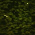
Fig. 8.3
Lymph node metastasis of synovial sarcoma. Axial contrast-enhanced CT image of the pelvis obtained for initial staging depicts enlarged external iliac lymph nodes on the right. Biopsy verified lymph node metastasis (arrows) of known biphasic synovial sarcoma. The primary tumor was located above the right knee
8.2.7 Distant Metastasis
The presence of distant metastasis represents the strongest predictor of poor outcome in soft tissue sarcomas [22, 32, 36]. Distant metastasis occurs in up to 25 % of patients with the most common sites being the lung (75–80 %) followed by the bones (10 %) [39]. CT of the chest should routinely be performed in initial staging of all soft tissue sarcomas (Fig. 8.4). Synchronous lung metastasis in the absence of extrapulmonary disease is usually treated by chemotherapy and, if possible, consequent surgical resection of all residual lesions. The standard treatment for metachronous resectable lung metastasis is surgery, if there is no evidence of extrapulmonary disease and complete excision of all lesions is feasible [37]. In specific sarcoma subtypes with a known tendency to metastasize to bone, such as myxoid liposarcoma, whole-body MRI may be recommended to assess the entire skeletal system.

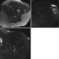
Fig. 8.4
Lung metastasis of alveolar soft part sarcoma. Axial CT image of the chest (lung window setting) obtained for initial staging shows bilateral pulmonary nodules (arrows). The primary tumor was located in the thigh
8.2.8 Histologic Grade
Histologic grading of soft tissue sarcomas according to the FNCLCC system is based on tumor differentiation, mitosis count, and tumor necrosis (Table 8.3) [7, 19]. The histologic grade has a higher prognostic weight than tumor size and depth [22]. It should be kept in mind that grading is not practicable in post-chemotherapy or post-radiotherapy specimens. It is therefore mandatory to obtain a sufficient amount of representative material for pathologic analysis prior to therapy.
Table 8.3
FNCLCC Grading System for Adult Soft Tissue Sarcomas [7] FNCLCC = Fédération Nationale des Centres de Lutte Contre le Cancer
Parameter | Criterion |
|---|---|
Tumor differentiation | |
Score 1 | Sarcomas closely resembling normal adult mesenchymal tissue |
Score 2 | Sarcomas for which histological typing is certain |
Score 3 | Embryonal sarcomas, undifferentiated sarcomas, and sarcomas of doubtful tumor type |
Mitosis count | |
Score 1 | 0–9 per 10 HPF |
Score 2 | 10–19 per 10 HPF |
Score 3 | >20 per 10 HPF |
Tumor necrosis | |
Score 0 | No necrosis
Related posts:Stay updated, free articles. Join our Telegram channel
Full access? Get Clinical Tree
 Get Clinical Tree app for offline access

|