The field of statistics makes valuable contributions to functional neuroimaging research by establishing procedures for the design and conduct of neuroimaging experiments and providing tools for objectively quantifying and measuring the strength of scientific evidence provided by the data. Two common functional neuroimaging research objectives include detecting brain regions that reveal task-related alterations in measured brain activity (activations) and identifying highly correlated brain regions that exhibit similar patterns of activity over time (functional connectivity). This article highlights various statistical procedures for analyzing data from activation studies and functional connectivity studies, focusing on functional magnetic resonance imaging (fMRI) and positron emission tomography (PET) data. Also discussed are emerging statistical methods for prediction using fMRI and PET data, which stand to increase the translational significance of functional neuroimaging data to clinical practice.
A range of substantive applications for functional neuroimaging methods exist, as other articles in this issue illustrate. For instance, functional neuroimaging has provided a better understanding of the neural basis for major psychiatric disorders, neurologic disorders, and substance abuse. Structural or anatomic neuroimaging, such as MRI, has had a tremendous impact on medical practice for both clinical diagnoses (eg, detecting brain lesions or tumors) and informing associated treatment decisions. Numerous functional neuroimaging studies, using functional MRI (fMRI) and cerebral blood flow positron emission tomography (PET), have identified neural processing differences or alterations in functional connectivity between patient groups and healthy controls, such as regarding Alzheimer’s disease . Although these group differences in neural processing suggest a role for functional neuroimaging in clinical practice, a paucity of research has targeted the development of standardized functional neuroimaging tools that aid in clinical decision making, especially in terms of treatment decisions. Nonetheless, important clinical contributions for functional neuroimaging are envisioned and this article discusses these possibilities.
The field of statistics has played an integral role in the advancement of functional neuroimaging research and has the potential to vastly increase the translational significance of neuroimaging techniques in clinical practice. Current functional neuroimaging research focuses largely on activation studies and studies of functional connectivity. Activation studies generally seek to characterize the neural responses to experimental tasks, which may be visualized as maps of distributed patterns of brain activity. Other common goals in activation studies are to detect differences in patterns of brain activity among various experimental stimuli, different subgroups of subjects, and two or more sessions (eg, before and after treatment). Whereas activation studies focus on localized brain activation, functional connectivity studies attempt to determine multiple brain areas that exhibit similar temporal task-related (or resting state) activity profiles. This article presents statistical methodology for analyzing functional neuroimaging data in activation studies and studies of functional connectivity. Furthermore, statistical techniques are described that show promise for clinical applications of functional neuroimaging data, enabling prediction of behavior-related neural processing, neural response to treatment, and clinical symptom response to treatment.
Statistical procedures for fMRI and PET data are highlighted that target activation studies, functional connectivity studies, and predictive techniques; a comprehensive review of statistical methods for functional neuroimaging data is beyond the scope of this article. To facilitate the presentation, the statistical methods are discussed in the context of fMRI data from a study of inhibitory control among cocaine-dependent men and PET data from a study of working memory in patients who have schizophrenia. General characteristics of fMRI and PET data are also briefly summarized.
Description of experimental data
Inhibitory Control in Cocaine-Dependent Men
Impairment in the ability to exert inhibitory control (eg, drug-seeking behaviors) despite adverse consequences, represents a common characteristic of drug addiction . The deficits in response inhibition that cocaine addicts often exhibit may be generalized outside of a drug-seeking context. The authors consider an fMRI study that evaluates the effects of cocaine addiction and treatment-related cocaine abstinence on the neural representation of inhibitory control experimental tasks. The cocaine-dependent subjects all enrolled in an addiction treatment program.
Baseline fMRI scans were collected from 41 men, 24 of whom were classified as cocaine-dependent and 17 as cocaine-free. Scans were obtained while subjects performed a variation of the classic Go/No-Go task, a stop task, and a Stroop task, all of which are designed to assess inhibitory control processes. The study reassessed neural processing related to the inhibitory control tasks 4 weeks after the initiation of treatment and tracked the time to relapse weekly for each subject for 1 year. This article considers data from the stop task.
General Characteristics of Functional MRI and Positron Emission Tomography Data
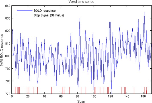
Functional neuroimaging using fMRI and PET measures hemodynamic correlates of localized brain activity. Each scan contains brain activity measurements at various spatial locations called voxels that are small cubic regions of roughly a few millimeters in size. The number of voxels in an image depends on the scan resolution, but as an example, a typical three-dimensional (3D) scan may consist of 2 mm 3 voxels configured in a 79 × 95 × 69 array after initial image preprocessing. The authors usually collect a series of 3D scans for each individual, obtained under different experimental conditions. The series of 3D scans represents a time series of image intensities at each voxel location, as depicted in Fig. 1 .
Related posts:
 Imaging of the Mind: Yesterday’s Triumphs and Tomorrow’s Challenges
Understanding Autism and Related Disorders: What has Imaging Taught Us?
Neuroimaging in Posttraumatic Stress Disorder and Other Stress-Related Disorders
Mood Disorders
The Role of Imaging in United States Courtrooms
Imaging of the Mind: Yesterday’s Triumphs and Tomorrow’s Challenges
Understanding Autism and Related Disorders: What has Imaging Taught Us?
Neuroimaging in Posttraumatic Stress Disorder and Other Stress-Related Disorders
Mood Disorders
The Role of Imaging in United States Courtrooms
 Visualizing White Matter Pathways in the Living Human Brain: Diffusion Tensor Imaging and Beyond
Stay updated, free articles. Join our Telegram channel

Full access? Get Clinical Tree


