36 Superficial Palmar Arch
B. Meyer, L. Sonnow
It is often difficult to distinguish between the types of the superficial palmar arch shown in Sections 36.1 and 36.2: the anastomosis can be so variable in size that the relative contribution to the supply of the hand is difficult to assess.
The number of types of anomalies actually observed is even larger than shown in these figures, since all four common palmar digital arteries do not originate from the superficial palmar arch (Chapter 37). Quite often, the first common palmar digital artery is absent, which normally supplies the ulnar side of the thumb and the radial side of the index finger. In such cases, the princeps pollicis artery is the only artery for that region. Because of the frequent absence of the first common palmar digital artery, only the next artery is sometimes designated the common palmar digital artery. This is called II in our figures because it is found in the second interosseous space.
In these figures, no differentiation has been made as to whether the feeding median artery is a “normal” median artery or a superficial median artery in the forearm.1–16
36.1 Closed Arch (42%)

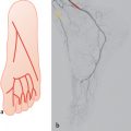
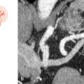
Fig. 36.1 “Normal” situation as described in textbooks (radioulnar type) (35%). Schematic (a) and DSA (b). 1 Deep palmar arch; 2 radial artery; 3 ulnar artery; 4 superficial palmar arch.

Fig. 36.2 Medianoulnar type (4%). Schematic (a) and DSA (b). 1 Radial artery; 2 interosseous artery; 3 median artery; 4 ulnar artery; 5 superficial palmar arch; 6 deep palmar arch.

Fig. 36.4 Profundoulnar type (the branch of the radial artery comes from the posterior side or from the deep palmar arch) (2%). Schematic (a), unsubtracted DSA (branch from the deep palmar arch) (b), and contrast-enhanced MR angiography, MIP (c). 1 Radial artery; 2 ulnar artery; 3 superficial palmar arch; 4 branch from the deep palmar arch; 5 incomplete deep palmar arch.
Stay updated, free articles. Join our Telegram channel

Full access? Get Clinical Tree


