Superficial Peroneal Nerve Block

48 Superficial Peroneal Nerve Block


The superficial peroneal nerve is a branch of the common peroneal nerve that emerges from the neck of the fibula between the extensor digitorum longus and peroneal muscles to enter the subcutaneous tissue of the lateral leg. When emerging from the fibular neck, the superficial peroneal nerve most commonly lies in the lateral compartment of the leg. The superficial peroneal nerve ascends along the anterior intermuscular septum to pierce the fascia lata at the juncture of the middle and lower thirds of the leg. The nerve usually divides into its medial and lateral branches once in the subcutaneous tissue.1,2


Some elect to infiltrate local anesthetic over the dorsum of the foot for superficial peroneal nerve block and reserve ultrasound for the deeper nerves of the ankle block.3 However, superficial peroneal nerve block in the leg can be useful when edema or infection contraindicates more distal ankle block. Proximal ultrasound-guided superficial peroneal nerve block (along with sural block) can provide surgical anesthesia for hardware removal from the lateral ankle in weight-bearing patients. In addition, ultrasound-guided superficial block in the leg is less painful than subcutaneous infiltration across the dorsum of the foot for more distal block and does not pierce the extensor tendons of the foot.
Suggested Technique


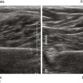
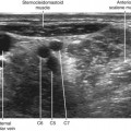
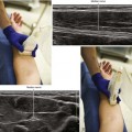
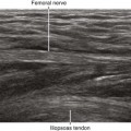
The superficial peroneal nerve can be difficult to image within the subcutaneous tissue of the distal lateral leg. By sliding the transducer along the known course of the nerve, the nerve can be identified as it emerges from the muscular compartment. An in-plane approach from either the anteromedial or posterolateral side of the leg can then be used for needle tip placement adjacent to the nerve.


More proximally, the peroneal nerves are best imaged in the prone position with the knee flexed using a pillow under the ankle. Alternatively, the leg can be elevated and internally rotated with the patient in supine position. When scanning from distal to proximal, the superficial peroneal nerve tracks along the fascia between the anterior and lateral compartments of the leg (like going down a ski jump), until it meets the acute edge of the bone of the fibula. The anterior border of the fibula is formed by the insertion of the anterior intermuscular septum. Because the nerve tracks along the anterior intermuscular septum, the anterior border of the fibula points toward the superficial peroneal nerve.


Because the superficial peroneal nerve most often lies in the lateral compartment side of the intermuscular septum, it is important for the block needle tip to puncture the septum when approaching from the anterior side. The block needle tip crosses the anterior intermuscular septum under the nerve, with injection of local anesthetic as the needle is pulled back. If the nerve is blocked close to the surface of the fascia lata, its deeper motor branches to the peroneus longus and peroneus brevis can be spared.

Stay updated, free articles. Join our Telegram channel

Mar 5, 2016 | Posted by in ULTRASONOGRAPHY | Comments Off on Superficial Peroneal Nerve Block

Full access? Get Clinical Tree

Get Clinical Tree app for offline access