58 Superior Laryngeal Nerve Block
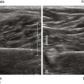
The superior laryngeal nerve lies close to the superior laryngeal artery. The internal branch of the superior laryngeal nerve lies on average 2.4 mm inferior to the greater horn of the hyoid bone.1 The hyoid bone is U shaped in transverse scans of the neck. The superior laryngeal nerve enters the larynx through an aperture (ostium) in the thyrohyoid membrane together with the superior laryngeal artery and vein.
Suggested Technique
Clinical Pearls
• The external branch of the superior laryngeal nerve lies near the superior pole of the thyroid gland. It can be difficult to identify, even with direct dissection and nerve stimulation.2
• The internal branch of the superior laryngeal nerve lies immediately inferior and deep to the greater cornu of the hyoid bone.3
Related posts:


Stay updated, free articles. Join our Telegram channel

Full access? Get Clinical Tree


