19 Superior Mesenteric Artery and Celiac Trunk
K.I. Ringe
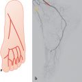
The basis for these figures are the anomalies of the celiac trunk and the hepatic arteries (for references, see Chapters 13 and 14). The variability of the gastroduodenal artery has also been considered.1–10 The combination of different accessory arteries has not been included. The figures in this chapters show that there is no clear-cut watershed between organs supplied by the celiac trunk and those supplied by the artery of the embryological midgut, the superior mesenteric artery.
19.1 Blood Supply of the Superior Mesenteric Artery to Abdominal Organs
In the drawings in this chapter, the following designations are used:
– Blank pattern = areas primarily supplied by branches of the celiac trunk.
– Dotted pattern = areas primarily supplied by the superior mesenteric artery, partly by branches of the celiac trunk.
– Striped pattern = areas primarily supplied by the superior mesenteric artery.

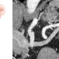
Fig. 19.1 “Normal” type as shown in textbooks (70%). Schematic (a) and coronal oblique 3D VR CT (b) with typical branching of the celiac trunk and superior mesenteric artery. 1 Common hepatic artery; 2 left gastric artery; 3 celiac trunk; 4 superior mesenteric artery; 5 splenic artery.

Fig. 19.2 Gastroduodenal artery from the superior mesenteric artery (5%). Schematic.

Fig. 19.3 Accessory right hepatic artery from the superior mesenteric artery (6%). Schematic (a) and coronal oblique 3D VR CT (b) in a patient with aortic dissection. 1 Right hepatic artery; 2 left hepatic artery; 3 common hepatic artery; 4 celiac trunk; 5 splenic artery; 6 right renal artery; 7 superior mesenteric artery; 8 accessory right hepatic artery.
Fig. 19.4 Right hepatic artery from the superior mesenteric artery (10%). Schematic (a) and subtracted DSA, with the diagnostic catheter placed in the superior mesenteric artery (b) and the celiac trunk (c). Patient with hepatocellular carcinoma (*). 1 Right hepatic artery; 2 superior mesenteric artery; 3 gastroduodenal artery; 4 left hepatic artery; 5 common hepatic artery; 6 splenic artery; 7 celiac trunk.
Stay updated, free articles. Join our Telegram channel

Full access? Get Clinical Tree


