Sural Nerve Block

49 Sural Nerve Block


The sural nerve forms from branches of the common peroneal (lateral) and tibial (medial) components of the sciatic nerve. Although the sural usually receives both contributions, anatomic variation in its composition is common.1


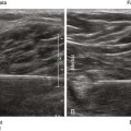
The sural nerve lies adjacent to the small saphenous vein within the subcutaneous tissue of the lateral leg. The sural nerve provides sensory innervation to the lateral foot.2 Because it is a sensory nerve, the sural nerve is sometimes used for biopsy or harvest. Although subcutaneous infiltration is an effective means of blocking the distal sural nerve, more proximal block may be indicated in patients with infection or edema of the foot.


Suggested Technique


Because of its small size, the sural nerve can be difficult to image. The sural nerve can be blocked proximal to the lateral malleolus by applying a calf tourniquet to help identify the small saphenous vein. In this location the sural nerve lies adjacent to the vein within the subcutaneous tissue of the leg. An in-plane approach can be used to distribute local anesthetic around the sural nerve.


A sural contribution from the tibial nerve can often be imaged between the medial and lateral heads of the gastrocnemius muscle. This contribution emerges between these muscles to pierce the fascia lata and join the lesser saphenous vein and the common peroneal nerve contribution within the subcutaneous tissue of the lateral aspect of the lower leg. The sural nerve can be blocked with an in-plane approach from the lateral aspect of the leg with the patient in supine position and the leg elevated. Although prone position is optimal for sural nerve imaging, the former approach is more practical and useful in most patients.


Key Points


Only gold members can continue reading. Log In or Register to continue

Stay updated, free articles. Join our Telegram channel

Mar 5, 2016 | Posted by in ULTRASONOGRAPHY | Comments Off on Sural Nerve Block

Full access? Get Clinical Tree

Get Clinical Tree app for offline access
Sural Nerve Block The Essentials
Anatomy The SuN emerges between the medial and lateral heads of the GC.
The SuN lies adjacent to the SSV within SQ tissue of the lateral leg.
The SuN is about 2.5 mm in diameter.