CHAPTER 9 Surface Anatomy of the Cerebrum
DESCRIPTIONS OF THE BRAIN SURFACE AND LABELING CODES USED IN THIS CHAPTER
Anterior parolfactory sulcus = subcallosal sulcus
Central sulcus = fissure of Rolando, rolandic fissure
Inferior frontal gyrus = triangular gyrus
Intraparietal sulcus = interparietal sulcus
Lingual gyrus = medial occipitotemporal gyrus (MOTG)
Lateral occipitotemporal gyrus (LOTG) = overlaps with fusiform gyrus
Middle occipital sulcus = lateral occipital sulcus, prelunate sulcus
Preoccipital notch = temporo-occipital incisure, temporo-occipital arcus
A separate nomenclature is also used for the gyri of the surface.
Pa: Ascending parietal gyrus (postcentral gyrus)
O4: Posterior intraoccipital portion of the LOTG
O5: Lingual gyrus (medial occipital-temporal gyrus)
T4: Anterior intratemporal portion of the LOTG
1 = superior frontal gyrus, F1
3 = inferior frontal gyrus, F3
4 = precentral gyrus, PreCG, P
5 = postcentral gyrus, PostCG, p
8 = superior parietal lobule, SPL
10 = superior temporal gyrus, T1
11 = middle temporal gyrus, T2
12 = inferior temporal gyrus, T3
13 = anterior intratemporal portion of the lateral occipitotemporal gyrus, T4, LOTG
14 = parahippocamapal gyrus, T5
15 = superior occipital gyrus, O1
16 = middle occipital gyrus, O2
17 = inferior occipital gyrus, O3
18 = posterior intraoccipital portion of the lateral occipitotemporal gyrus, O4, LOTG
19 = lingual gyrus (medial occipitotemporal gyrus), O5, MOTG
20 = gyrus descendens (of Ecker)
31 = isthmus of the cingulate gyrus
36 = lateral occipitotemporal gyrus, LOTG
a = superior frontal sulcus, SFS
b = inferior frontal sulcus, IFS
e = postcentral sulcus, postCS
g = intra-occipital sulcus, IOS
h = lateral (middle) occipital sulcus
i = transverse occipital sulcus
k = parieto-occipital sulcus, POS
p = superior temporal sulcus, STS
q = inferior temporal sulcus, ITS
v = pars marginalis of cingulate sulcus
y = intra-occipital sulcus, IOS
The surface of the cerebrum is typically subdivided into lateral (convexity), medial, superior, and inferior surfaces, separated by angular edges designated margins.1–3 The brain surface forms a continuous sheet of tissue that is folded and pleated to variable depths to form outwardly directed folds (the gyri), inwardly directed folds (the sulci), and large overhanging lips (the opercula) that cover the insula of each side. Individual gyri and sulci commonly continue from one surface across a margin onto an adjacent surface. The sulcal pattern shows wide variation among individuals, variation from side to side, and variation with patient handedness and language dominance (see section on petalia).1 Overall, however, the pattern conforms to recognizable ranges of variation that permit ready identification of the sulci and gyri in most individuals.1
EMBRYOLOGY
The gyri and sulci mature in a reproducible pattern from fetal to infantile to adult age (Fig. 9-1).4–9 Sulci first appear as linear depressions in the smooth brain surface (primary sulci). The primary sulci lengthen and deepen with time but retain relatively simple linear and curvilinear shapes. With maturation, the ends of the primary sulci typically bifurcate to form secondary sulci. These secondary sulci may later bifurcate to form tertiary sulci. The additional folds give the brain surface an increasingly complex appearance with age (see Fig. 9-1). Appreciation of this maturation pattern makes it simpler to understand the more complex folds, and variations, of the adult.9–25
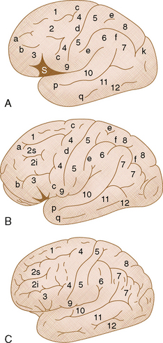
FIGURE 9-1 Maturation of the convexity sulci. Diagrammatic representations. Gyri: 1, Superior frontal gyrus; 2, middle frontal gyrus; 3, inferior frontal gyrus; 4, precentral gyrus; 5, postcentral gyrus; 6+7, inferior parietal lobule composed of the supramarginal gyrus (6) and the angular gyrus (7); 8, superior parietal lobule; 9, subcentral gyrus; 10, superior temporal gyrus; 11, middle temporal gyrus; 12, inferior temporal gyrus. A, At approximately 7 to 9 months’ gestation. The opercula have not yet completed their folding, so the sylvian fissure (S) is widely open. The superior frontal sulcus (a) and inferior frontal sulcus (b) begin as a series of shallow longitudinal depressions on the convexity surface. The posterior ends of these sulci bifurcate forming separate upper and lower segments of the precentral sulcus (c). The central sulcus (d) is isolated from the sylvian fissure by the subcentral gyrus (9). The postcentral sulcus (e) also forms from two separate segments. The lower postcentral segment is simultaneously the upswing of the arcuate intraparietal sulcus (f). This sulcus presently describes a simple arc with little definition of the individual supramarginal (6) and angular (7) gyri. The parieto-occipital sulcus (k) is well developed. On the temporal convexity, the superior temporal sulcus (p) is best developed early. The inferior temporal sulcus (q) is immature. B, At approximately birth to 2 years of age. The opercula have closed together, narrowing the sylvian fissure. The individual segments of the superior (a) and inferior (b) frontal sulci have merged into unified lengths. The large middle frontal gyrus (2) is partially subdivided into superior and inferior segments (2s and 2i) by the middle frontal sulcus (not labeled). The posterior bifurcations of the superior and inferior frontal sulci form two separate segments of the precentral sulcus (c). These two segments do not merge together, leaving a gap through which the posterior portion of the middle frontal gyrus (2) unites with (was never separated from) the anterior face of the precentral gyrus (4). The intraparietal sulcus (f) is now better developed and lobulated, defining the supramarginal (6) and angular (7) gyri. The inferior temporal sulcus (q) has matured with greater length of its segments. C, After approximately age 2 years. With greater maturation, the sulci lengthen, deepen, and become deflected by the growth of the neighboring gyri. Their ends bifurcate to form secondary sulci, and the bifid ends bifurcate again to form tertiary sulci. Additional local folding creates unnamed folds over the surfaces of the named gyri. Further details are available in references 8 and 9.
(Based on data from Turner OA. Growth and development of the cerebral cortical pattern in man. Arch Neurol Psychiatry 1948; 59:1-12.)
ANATOMY
Lateral Surface
The lateral surface of the cerebrum includes the entire C-shaped convexity of the brain that extends around the sylvian fissure from the frontal pole anteriorly to the occipital pole posteriorly to the temporal pole inferiorly (Figs. 9-2 and 9-3).14,20,25 The lateral surface is subdivided into lobes by prominent intrinsic landmarks such as the central sulcus, sylvian fissure, and parieto-occipital sulcus; inconstant “landmarks” such as the preoccipital notch; and arbitrary lines including the lateral parietotemporal line and the temporo-occipital line (see Figs. 9-3 and 9-4).1 The lateral parietotemporal line is drawn from the lateral end of the parieto-occipital sulcus superiorly to the preoccipital notch inferiorly. The temporo-occipital line is drawn from the posterior end of the sylvian fissure to the midpoint of the lateral parietotemporal line. The lateral surface of the cerebrum contains portions of the frontal, parietal, occipital, and temporal lobes, arrayed around the sylvian (lateral) fissure. With the just-noted landmarks, the lateral surface of the frontal lobe extends from the frontal pole to the central sulcus above the sylvian fissure. The lateral surface of the parietal lobe extends from the central sulcus to the parietotemporal line above the sylvian fissure and above the temporo-occipital line. The lateral surface of the temporal lobe extends from the temporal pole to the lateral parietotemporal line inferior to both the sylvian fissure and the temporo-occipital line. The lateral surface of the occipital lobe extends from the lateral parietotemporal line to the occipital pole. Because the occipital lobe curves sharply medially toward the occipital pole, true lateral views give a deceptively foreshortened impression of the size of the lateral occipital surface.
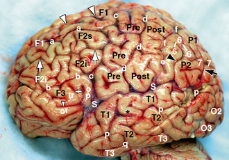
FIGURE 9-2 Surface features of the left hemisphere. Fresh gross specimen with pia-arachnoid and vessels intact. The convexity surface of the hemisphere displays four frontal gyri: the superior frontal gyrus (F1), middle frontal gyrus (F2), inferior frontal gyrus (F3), and the precentral gyrus (Pre); three temporal gyri: the superior temporal gyrus (T1), middle temporal gyrus (T2), and inferior temporal gyrus (T3); three subdivisions of the parietal lobe: the postcentral gyrus (Post), superior parietal lobule (P1), and inferior parietal lobule (P2); and three subdivisions of the occipital lobe: the superior occipital gyrus (not seen from this view), middle occipital gyrus (O2), and the inferior occipital gyrus (O3). Short inconstant medial frontal sulci (large white arrowheads) often groove F1. A longitudinal middle frontal sulcus (large white arrows) commonly divides F2 into superior (F2s) and inferior portions (F2i). These halves may unite with the adjacent F1 and/or F3 gyri in complex ways. The anterior horizontal ramus (small white arrowhead) and the anterior ascending ramus (small white arrow) of the sylvian fissure (S) extend into F3, dividing it into the pars orbitalis (or), pars triangularis (tr), and pars opercularis (op). They give F3 the shape of an upper case M. The superior frontal sulcus (a) separates F1 from F2 below. The inferior frontal sulcus (b) separates F2 from F3 below. The posterior end of the superior frontal sulcus (a) characteristically bifurcates to form the superior precentral sulcus (c) that separates F1 from the upper precentral gyrus (Pre). The posterior end of the inferior frontal sulcus (b) characteristically bifurcates to form the inferior precentral sulcus (c) that separates F3 from the lower precentral gyrus (Pre). F2 characteristically unites with the anterior face of the precentral gyrus (Pre) between the upper (c) and lower (c) portions of the precentral sulcus. The central sulcus (d) separates the precentral gyrus from the postcentral gyrus. The central sulcus is usually isolated from the sylvian fissure (S) by a subcentral gyrus, but not in this case. The upper and lower portions of the postcentral sulcus (e) separate the postcentral gyrus from the superior parietal (P1) and inferior parietal (P2) lobules. The lower portion of the postcentral sulcus is, simultaneously, the ascending portion of the arcuate intraparietal sulcus (f) that separates the superior parietal lobule (8) from the inferior parietal lobules (6+7). The ascending ramus (black arrowhead) of the sylvian fissure (S) angles upward into the inferior parietal lobule. The horseshoe-shaped gyrus draped over the posterior ascending ramus of the sylvian fissure is the supramarginal gyrus (6). The superior temporal sulcus (p) separates the superior temporal gyrus (T1) from the middle temporal gyrus (T2). The inferior temporal sulcus (q) separates the middle temporal gyrus (T2) from the inferior temporal gyrus (T3). Anteriorly, a short vertical gyrus (black asterisk) commonly extends between T1 and T2, interrupting the superior temporal sulcus (p) over a short segment. Posteriorly, the superior temporal sulcus angles upward in parallel with the posterior ascending ramus of the sylvian fissure over a segment designated the angular sulcus (large black arrow). The horseshoe-shaped gyrus draped over the angular sulcus is the angular gyrus (7). Together the supramarginal and angular gyri constitute most of the inferior parietal lobule.
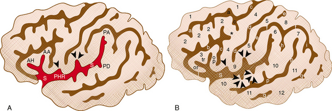
FIGURE 9-3 Convexity surface. Diagrammatic representation. A, The margins of the frontal, parietal and temporal opercula are defined by the sylvian fissure (S), by its five major rami (the anterior horizontal ramus [AH], anterior ascending ramus [AA], posterior horizontal ramus [PHR], posterior ascending ramus [PA] and posterior descending ramus [PD]) and by its minor arms (the anterior subcentral sulcus [single arrowhead], posterior subcentral sulcus [double arrowheads], and the transverse temporal sulci [triple arrowheads on B]). B, The configuration of the sylvian fissure then permits identification of the surface gyri and sulci. Gyri: 1, superior frontal; 2, middle frontal; 3, inferior frontal; 4, precentral; 5, postcentral; 6, supramarginal; 7, angular; 8, superior parietal lobule; 9, subcentral; 10, superior temporal, 11, middle temporal; 12, inferior temporal gyrus. Asterisk indicates junction of the middle frontal gyrus with the precentral gyrus. Sulci: a, superior frontal sulcus; b, inferior frontal sulcus; c, superior and inferior segments of the precentral sulcus; d, central sulcus; e, superior and inferior segments of the postcentral sulcus; f, intraparietal sulcus; p, superior temporal sulcus; q, inferior temporal sulcus; h, primary intermediate sulcus; i, secondary intermediate sulcus.
(Modified from Naidich TP, Valavanis AG, Kubik S, et al. Anatomic relationships along the low-middle convexity: II. Lesion localization. Int J Neuroradiol 1997; 3:393-409.)
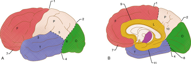
FIGURE 9-4 Lobar boundaries and nomenclature. A, Convexity (lateral) surface. On the convexity, the central sulcus (1) separates the frontal (F) from the parietal (P) lobes. The sylvian fissure (3) separates the frontal from the temporal (T) lobes. The demarcation of the temporal, parietal, and occipital lobes is arbitrary and inconstant from publication to publication. In one system, a parietotemporal line is drawn from the lateral edge of the parieto-occipital sulcus (2) to the preoccipital notch (temporo-occipital incisure) (4). This line sets the arbitrary anterior border of the occipital lobe (O), separating it from the parietal and temporal lobes anterior to it. A second arbitrary temporo-occipital line (5) is drawn from the posterior descending ramus of the sylvian fissure (3) to the middle of the parietotemporal line (6). This line sets the arbitrary parietotemporal boundary. B, Medial surface. On the medial surface, the central sulcus (1) usually curves onto the medial surface perpendicular to the marginal segment of the cingulate sulcus. A line drawn from the central sulcus to the cingulate sulcus establishes the frontoparietal border. The deep parieto-occipital sulcus (2) demarcates the parietal lobe from the occipital lobe. An arbitrary basal parietotemporal line (8) drawn from the inferior end of the parieto-occipital sulcus to the preoccipital notch establishes the temporal (T)/occipital (O) border. The limbic lobe (L) is delimited by the cingulate sulcus (9), the subparietal sulcus (10), and the collateral sulcus (11). Also labeled: 7, orbital surface.
Frontal Lobe
The convexity surface of the frontal lobe displays four major gyri (Figs. 9-2 to 9-6). Anteriorly, the longitudinally oriented superior frontal gyrus, middle frontal gyrus, and inferior frontal gyrus are separated from each other by the superior and inferior frontal sulci. The middle frontal gyrus is the largest of the three gyri and may be partially subdivided into upper and lower halves by an inconstant middle frontal sulcus. The convexity surface of the superior frontal gyrus may be grooved by short shallow sulci termed the medial frontal sulci. Posteriorly, the frontal lobe is formed by the precentral gyrus that courses vertically between the precentral sulcus anteriorly and the central sulcus posteriorly. The inferior end of the central sulcus usually does not intersect the sylvian fissure. Instead, a U-shaped subcentral gyrus is interposed between the inferior end of the central sulcus and the sylvian fissure.
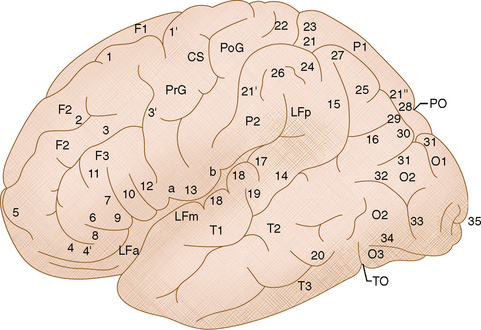
FIGURE 9-5 Surface features of the convexity. Diagrammatic representation.
Gyri: F1, F2, and F3, superior, middle, and inferior frontal gyri; P1 and P2, superior and inferior parietal lobules; T1, T2, and T3, superior, middle, and inferior temporal gyri; O1, O2, and O3, superior, middle, and inferior occipital gyri; PrG and PoG, precentral and postcentral gyri; PO, parieto-occipital fissure; TO, temporal-occipital incisure; LFa, LFm, and LFp, lateral fissure of Sylvius (anterior [a], middle [m] and posterior [p] segments).
Frontal lobe: 1, superior frontal sulcus; 1′, superior precentral sulcus; 2, inconstant middle frontal sulcus; 3, inferior frontal sulcus; 3′, inferior precentral sulcus; 4, lateral orbital sulcus; 4′, lateral orbital gyrus; 5, frontomarginal sulcus; 6, anterior horizontal ramus of the sylvian fissure; 7, anterior ascending ramus of the sylvian fissure; 8-10, partes orbitalis (8), triangularis (9), and opercularis (10) of the inferior frontal gyrus; 11, sulcus triangularis; 12, sulcus diagonalis within the pars opercularis; 13, subcentral gyrus delimited by the anterior (a) and posterior (b) subcentral sulci.
Temporal lobe: 14, superior temporal sulcus (parallel sulcus) anterior segment; 15, superior temporal sulcus, ascending posterior segment (synonym: angular sulcus); 16, superior temporal sulcus, horizontal posterior segment; 17, transverse temporal sulcus; 18, transverse temporal gyri; 19, sulcus acousticus; 20, inferior temporal sulcus.
Parietal lobe: 21, intraparietal sulcus, horizontal segment; 21′, intraparietal sulcus, ascending segment (coincident with inferior postcentral sulcus); 21″, intraparietal sulcus, descending segment; 22, superior postcentral sulcus; 23, transverse parietal sulcus; 24, primary intermediate sulcus (of Jensen); 25, secondary intermediate sulcus; 26, supramarginal gyrus; 27, angular gyrus; 28, first parieto-occipital arcus (first pli du passage of Gratiolet); 29, second parieto-occipital arcus (second pli du passage of Gratiolet).
Occipital lobe: 30, intraoccipital sulcus (superior occipital sulcus); 31, transverse occipital sulcus; 32, lateral (middle) occipital sulcus; 33, lunate sulcus; 34, inferior occipital sulcus; and 35, calcarine sulcus, here extending to the occipital pole.
(From Duvernoy HM. The Human Brain: Surface, Three-Dimensional Sectional Anatomy and MRI. New York, Springer, 1991.)
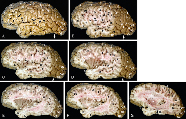
FIGURE 9-6 Convexity surface of the brain with sequential sagittal sections. Formalin-fixed gross anatomic specimen after removal of the pia-arachnoid and vessels (same specimen as Fig. 9-10). A and B, The sylvian fissure (S) separates the frontal and parietal lobes superiorly from the temporal lobe inferiorly. At its anterior end, the anterior horizontal (small black arrowhead) and anterior ascending (small black arrow) rami subdivide the inferior frontal lobe into partes orbitalis (or), triangularis (tr), and opercularis (op). Posteriorly, the posterior ascending ramus (most posterior of the three S) deeply indents the supramarginal gyrus (6), giving it a horseshoe shape. The posterior descending ramus of the sylvian fissure (two small black arrowheads) is typically very small. The convexity surface of the frontal lobe is composed of the 1, superior frontal gyrus; 2s and 2i, superior and inferior segments of the middle frontal gyrus; 3, M-shaped inferior frontal gyrus; and 4, precentral gyrus. These are delimited by the superior frontal sulcus (a), the inferior frontal sulcus (b), and the precentral sulcus (c). A vertical triangular sulcus grooves the pars triangularis (between the letters t and r). The superior surface of pars triangularis commonly unites with the inferior segment of the middle frontal gyrus (2i) across the inferior frontal sulcus (b). The convexity surface of the parietal lobe is composed of the 5, postcentral gyrus; 6-7, the inferior parietal lobule formed by the supramarginal gyrus (6) and angular gyrus (7); and 8, the superior parietal lobule. These are delimited by the central sulcus (d), postcentral sulcus, intraparietal sulcus (f), middle (lateral) occipital sulcus (h), and the transverse occipital sulcus (i). The subcentral gyrus (9) links the inferior ends of the precentral (5) and postcentral (6) gyri inferior to the central sulcus (d). The inferior end of the postcentral gyrus (5) shows a deep partition. The posterior portion may be considered an accessory presupramarginal gyrus (double asterisks). The inferior portion of the postcentral sulcus constitutes the ascending segment of the arcuate intraparietal sulcus (f). The transverse occipital sulcus (i) separates the parietal lobe (7) from the middle occipital gyrus (O2). Just as the large middle frontal gyrus (2) is commonly divided into upper and lower portions by a middle frontal sulcus, the large middle occipital gyrus (O2) is often separated into upper and lower portions by a middle (lateral) occipital sulcus (h). C, This section just exposes the anterior lobule of the insula and the relationship of the partes orbitalis, triangularis, and opercularis to the insula. D to F, The insula is delimited by a circular (peri-insular) sulcus. The larger anterior lobule of the insula has three (or more) short insular gyri: anterior short (as), middle short (ms), and posterior short (ps). These converge inferiorly to form the apex of the anterior lobule. The posterior lobe has two long insular gyri: anterior long (al) and posterior long (pl). The central sulcus of the convexity (c) continues across the insula (white dashes) between the anterior and posterior insular lobules. It then swings immediately under the apex to pass medially toward the suprasellar cistern. Heschl’s transverse temporal gyrus forms a distinct elevation (black plus sign) on the upper surface of the superior temporal gyrus. It characteristically arises immediately behind the posterior lobule of the insula and angles obliquely across the upper surface of the temporal lobe (see Fig. 9-7). G, This section cuts deep to the insula exposing the putamen (pu). Entry into the lateralmost portion of the temporal horn (two black arrows) exposes the lateral surface of the hippocampal formation that lies along the medial wall of the horn.
Parietal Lobe
The convexity surface of the parietal lobe displays three major subdivisions (see Figs. 9-2 to 9-6). Anteriorly, the vertically oriented postcentral gyrus lies between the central sulcus anteriorly and the postcentral sulcus posteriorly. Posteriorly, the deep, arcuate intraparietal sulcus subdivides the lateral surface of the parietal lobe into superior and inferior parietal lobules. The superior parietal lobule forms the superomedial portion of the parietal convexity between the superior margin of the hemisphere and the intraparietal sulcus. The inferior parietal lobule forms the inferolateral portion of the parietal convexity between the intraparietal sulcus and the temporo-occipital line. The inferior parietal lobule contains the supramarginal gyrus anteriorly and the angular gyrus posteriorly.
Occipital Lobe
The convexity surface of the occipital lobe displays three major gyri: the superior occipital gyrus, middle occipital gyrus, and inferior occipital gyrus, separated by the superior and inferior occipital sulci (see Figs. 9-3, 9-5, and 9-6). The superior occipital sulcus is usually seen as the posterior continuation of the intraparietal sulcus. The inferior occipital sulcus is usually seen as the posterior extension of the inferior temporal sulcus. The middle occipital gyrus is the largest of the three occipital gyri and may be partially subdivided into upper and lower halves by an inconstant middle (lateral) occipital sulcus. Far posteriorly, the convexity surface of the occipital lobe often displays a vertically oriented, arcuate lunate sulcus. The posterior end of the calcarine sulcus may extend around the occipital pole to lie on the convexity surface.
Temporal Lobe
The lateral surface of the temporal lobe displays three major gyri that course longitudinally inferior to the sylvian fissure and inferior to the temporo-occipital line (see Figs. 9-4, 9-5, and 9-6). The superior temporal, middle temporal, and inferior temporal gyri are separated by the superior and inferior temporal sulci. The transverse temporal gyrus of Heschl forms a focal protuberance on the superior surface of the superior temporal gyrus (Figs. 9-7 and 9-8). This deflects the sylvian fissure upward focally. The inferior temporal gyrus forms the inferior margin of the hemisphere and curves onto the inferior surface of the hemisphere to form the lateralmost gyrus of the inferior temporal surface (Fig. 9-9). The preoccipital notch (temporo-occipital incisure) marks the transition from temporal lobe to occipital lobe along the inferior margin (see Figs. 9-5 and 9-6).
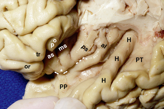
FIGURE 9-7 Superior temporal plane and the primary auditory cortex (H). Formalin-fixed gross anatomic specimen. Resection of the frontal lobe posterior to the inferior frontal gyrus opens a view of the upper surface of the temporal lobe, designated the superior temporal plane, the transverse temporal gyrus of Heschl (H), Heschl’s sulcus immediately behind the gyrus, and two broad flat planes of tissue anterior and posterior to Heschl’s gyrus and sulcus. From the temporal pole anteriorly to the front of Heschl’s gyrus, the flat surface is designated the planum polare (PP). From Heschl’s sulcus to the posterior end of the temporal surface, the flat surface is designated the planum temporale (PT). The planum temporale is usually triangular, with its point medial and its base directed laterally. It is usually larger in the language-dominant temporal lobe. Note that Heschl’s gyrus commonly bifurcates at its lateral end. The partes orbitalis (or), triangularis (tr), and opercularis (op) of the inferior frontal gyrus overhang the anterior lobule of the insula. The anterior lobule displays the anterior short (as), middle short (ms), and posterior short (ps) gyri. These converge to the apex (asterisk) of the insula inferiorly. The central sulcus of the insula (dashed white lines) separates the anterior lobe from the posterior lobe of the insula and then swings medially under the apex toward the suprasellar cistern.
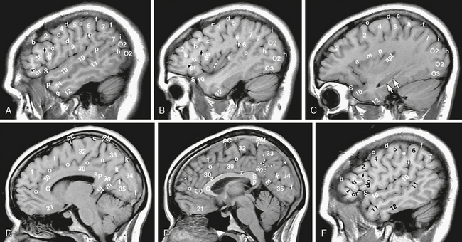
FIGURE 9-8 A to C and F, Lateral surface T1W MR images (labels as in Fig. 9-6). D, E, Medial surface T1W MR images (labels as in Fig. 9-10). MRI displays all of the surface features seen by gross inspection of the brain. The contralateral side of this same patient (see F) illustrates a common variation of the inferior frontal gyrus. Here, the anterior ascending ramus (small black arrow) of the sylvian fissure cuts completely through the gyrus, leaving pars opercularis as an anterior appendage of the lower precentral gyrus (4). Pars triangularis bridges across the inferior frontal sulcus to join the inferior portion of the middle frontal gyrus on the next section (not shown). In C, amp, short insular gyri; apl, long insular gyri.
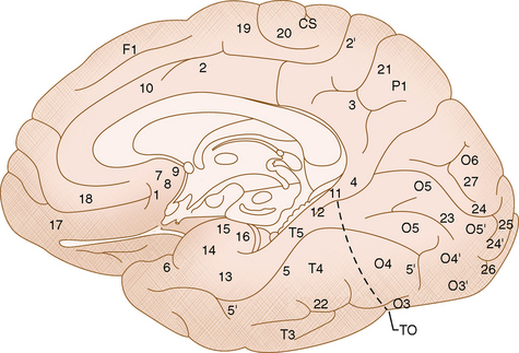
FIGURE 9-9 Surface features of the inferomedial brain. Diagrammatic representation.
Gyri: T3, T4, and T5, inferior temporal gyrus, temporal portion of the fusiform gyrus, parahippocampal gyrus; respectively; P1, medial aspect of the superior parietal lobule (precuneus); O3, O4, and O5, inferior occipital gyrus, occipital portion of the fusiform gyrus, lingual (medial occipitotemporal gyrus), respectively; O3 plus O4′ plus O5′, the caudal portions of O3, O4, and O5 merge into a common occipital cortex on the inferior aspect of the occipital pole. T4 plus O4 form the fusiform (lateral occipitotemporal) gyrus. TO, Temporo-occipital incisure. The limbic lobe is delimited, sequentially, by the 1, anterior parolfactory sulcus (subcallosal sulcus); 2, cingulate sulcus; 3, subparietal sulcus; 4, anterior calcarine sulcus; 5, collateral sulcus (medial occipitotemporal sulcus); and 6, rhinal sulcus. The limbic lobe is composed, sequentially, of the 7, subcallosal area; 8, posterior parolfactory sulcus; 9, paraterminal gyrus; 10, cingulate gyrus; 11, isthmus of the cingulate gyrus; 12, dentate gyrus; and 13, pyriform lobe (itself formed of the 14, gyrus ambiens; 15, semilunar gyrus; and 16, limbus [band] of Giacomini). Also labeled: 2′, marginal segment (pars marginalis) of the cingulate sulcus, and 5′, anterior and posterior transverse collateral sulci.
Medial frontal lobe: 17, gyrus rectus; 18, supraorbital sulcus; 19, paracentral sulcus; 20, paracentral lobule; CS, superior end of the central sulcus on the medial surface.
Medial parietal lobe: 21, transverse parietal sulcus.
Inferomedial temporal lobe: 22, lateral occipitotemporal sulcus.
Inferomedial occipital lobe: 23, inconstant lingual sulcus (when present it divides the lingual gyrus into upper and lower portions); 24, calcarine sulcus; 24′, retrocalcarine cortex; 25, gyrus descendens; 26, occipitopolar sulcus; 27, paracalcarine sulcus.
(From Duvernoy HM. The Human Brain: Surface, Three-Dimensional Sectional Anatomy and MRI. New York, Springer, 1991.)
Opening the sylvian fissure discloses the superior surface of the temporal lobe, termed the superior temporal plane. This forms the inferior lip (temporal operculum) of the sylvian fissure. The transverse temporal gyrus of Heschl arises posteromedially immediately behind the insula and courses anterolaterally across the superior temporal plane to reach the lateral surface. Heschl’s sulcus defines its posterior border (see Figs. 9-6 and 9-7). The portion of the superior temporal plane that lies between the temporal pole and the anterior surface of Heschl’s gyrus is termed the planum polare. This is delimited medially by the circular sulcus of the insula (see later). The portion of the superior temporal plane that lies behind Heschl’s gyrus and sulcus is termed the planum temporale. Because of the oblique course of Heschl’s gyrus, the planum temporale appears triangular, with its base on the convexity surface and its apex directed posteromedially, immediately behind the origin of Heschl’s gyrus. The planum temporale is usually larger on the side of language dominance.
Insular Lobe
Opening the lips of the sylvian fissure discloses the insula (synonym: island of Reil) (see Figs. 9-6 and 9-7).25 The insula is circumscribed by the circular (peri-insular) sulcus, which is subdivided into anterior, superior, and inferior peri-insular segments. The central sulcus of the convexity extends onto the insula, crosses the insular surface between the anterior insular lobule and the posterior insular lobule, and then curves medially into the deep sylvian fissure. The larger anterior insular lobule commonly displays three vertically oriented gyri referred to as the anterior short, middle short, and posterior short insular gyri. Additional inconstant anterior insular gyri are common. Typically, the inferior ends of the anterior, middle, and posterior short insular gyri converge together to form the apex of the insula. The central sulcus courses immediately beneath the apex as it turns medially into the deep sylvian fissure. The anterior insular lobule connects directly with the posteromedial orbital lobule on the orbital surface of the frontal lobe (see Inferior Surface). The smaller posterior insular lobule typically displays two obliquely oriented gyri: the anterior long and posterior long insular gyri. In some ways, the transverse temporal gyrus of Heschl may be regarded as a third long insular gyrus that happened to be folded onto the superior surface of the temporal lobe by the development of the temporal operculum.
Central Lobe
The tissue surrounding the central sulcus has been proposed to form a separate lobe, referred to as the central lobe.21 The tissue surrounding the central sulcus does form a continuous loop that can be followed, round and round, from the precentral gyrus anteriorly into the subcentral gyrus inferiorly, into the postcentral gyrus posteriorly into the paracentral lobule superiorly, and then back into the precentral gyrus, and so on. Because the pericentral gyri subserve sensorimotor function, the concept of a separate central lobe merits consideration, although it is not yet widely accepted.
Medial Surface
Like the lateral surface, the medial surface of the cerebrum is divided into lobes by prominent intrinsic landmarks such as the cingulate sulcus, central sulcus, parieto-occipital sulcus, and collateral sulcus; inconstant “landmarks” such as the preoccipital notch; and an arbitrary basal parietotemporal line drawn from the inferior end of the parieto-occipital sulcus superiorly to the preoccipital notch inferiorly.1,2,17 Unlike the lateral surface, the gyri and sulci of the medial surface are arranged in a radial coordinate system, in which all of the gyri form arcs of tissue that course co-curvilinear with the corpus callosum or perpendicular (radial) to the curvature of the corpus callosum. The central landmark of the medial surface is the corpus callosum, subdivided into the rostrum anteroinferiorly, the genu anteriorly, the body superiorly, and the splenium posteriorly (see Figs. 9-4 and 9-9). Grossly, the outer margin of the corpus callosum is delimited by the callosal sulcus Actually, however, the external surface of the corpus callosum is covered by a thin layer of gray matter: the indusium griseum superiorly and the paraterminal gyrus anteroinferiorly.
Limbic Lobe
The limbic lobe is the name given to that portion of the medial surface of the hemisphere that encircles the corpus callosum and extends along the superomedial surface of the temporal lobe to encircle the brain stem (Fig. 9-10
Stay updated, free articles. Join our Telegram channel

Full access? Get Clinical Tree


