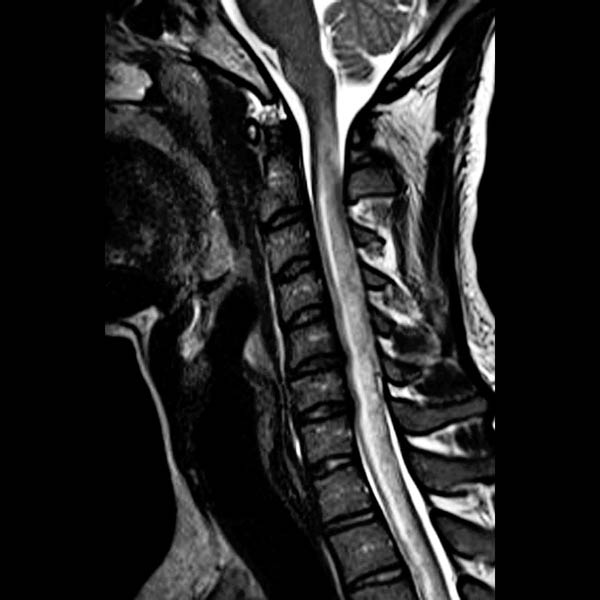
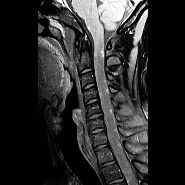
(Left) Sagittal T2WI MR shows diffuse cord edema as T2 hyperintensity with diffuse cord expansion obscuring any CSF surrounding cervical cord. (Courtesy E. Gasparetto, MD.)
(Right) Sagittal T1WI C+ FS MR shows multiple nodular foci of enhancement primarily located on the cord pial surface extending from medulla throughout the cervical cord. More severe pial involvement is referred to as “candle guttering” pattern with linear and nodular foci. (Courtesy E. Gasparetto, MD.)
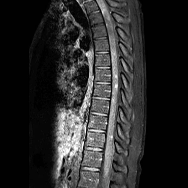
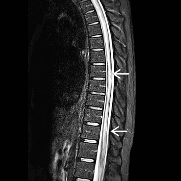
(Left) Sagittal STIR MR shows diffuse T2 hyperintensity throughout the thoracic cord in this patient with subacute syphilitic myelitis. Slight irregularity or T2 hyperintensity corresponds to foci of enhancement. (Courtesy E. Gasparetto, MD.)
(Right) Sagittal T1WI C+ FS MR shows multiple nodular foci of enhancement throughout the thoracic cord, with a strong predominance toward the cord pial surface. (Courtesy E. Gasparetto, MD.)
TERMINOLOGY
Synonyms
• Neurosyphilis, syphilitic meningomyelitis
Definitions
• CNS infection by spirochete Treponema pallidum
IMAGING
General Features
• Best diagnostic clue
Neurosyphilis diagnosis requires 1 of the following CSF pathologies
– Increased cell count or protein
– Reactive VDRL or RPR
– Positive iTPA index
– Positive PCR for T.pallidum
• Location
Pial surface of cord &/or intramedullary
• Size
Variable nodular and linear enhancement
Imaging Recommendations
• Best imaging tool
Enhanced MR to define cord edema and enhancing leptomeningeal or cord parenchymal enhancement
Only gold members can continue reading. Log In or Register to continue

 corresponds to foci of enhancement. (Courtesy E. Gasparetto, MD.)