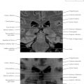
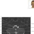
Temporal Bone Axial 1

Normal Anatomy
The temporal bone initially may seem a daunting area for magnetic resonance imaging because of the apparent structural complexity. To understand the temporal region better, trace the hearing pathway from auricle through external auditory canal, to tympanic membrane attached to small bones (ossicles; malleus, incus, and stapes) in the middle ear cavity, through stapes stirrup on oval window, then vestibule to cochlea to inner auditory canal (IAC). The vestibulocochlear nerve (cranial nerve [CN] VIII) also provides balance with superior and inferior vestibular branches in the IAC, leading to the semicircular canals. Finally, the nearby facial nerve (CN VII) provides motor innervation to the face.
These nerves can be tracked easily on computed tomography (CT) by the corresponding bony canals, although the nerves themselves are best seen on MRI (see also Chapter 5 ).

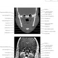
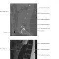
Temporal Bone Axial 2

Pathologic Process
Note the fan-shaped appearance of the labyrinthine segment of the facial nerve connecting the intracanalicular segment with the geniculate ganglion more anteriorly. Damage to the facial nerve, as occurs with trauma and temporal bone fracture, leads to loss of motor control on the ipsilateral side of the face, mimicking a stroke, with profound social impact given the sensitivity to facial expression. (See facial nerve [CN VII] images in Chapter 5 .)

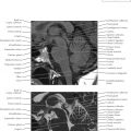
Temporal Bone Axial 3

Normal Anatomy
Note the apical and middle turns of the cochlea. Counterintuitively, the tip of the cochlea (the apex) is sensitive to lower frequencies, down to 20 Hz, moving to higher frequencies in the middle turn, and up to 20,000 Hz in the basal turn.
Note also the very thin vestibular aqueduct, which connects the inner ear to a balloon-shaped structure called the endolymphatic sac. Although its function is not clearly known, the endolymphatic sac may help to ensure that fluid in the inner ear contains the correct ion concentration. The vestibular aqueduct is normally less than 1 mm in diameter, or smaller than the adjacent medial limb of the superior semicircular canal (a good anatomic reference). Enlarged vestibular aqueducts, associated with hearing loss in children, are thought to be related to the same underlying defect.
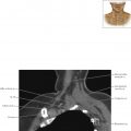
Pathologic Process
Damage to the cochlear nerve, as in trauma with temporal bone fracture, is more classically seen with transverse fractures perpendicular to the long axis of the temporal bone, from auricle to petrous apex. Transverse fractures are more frequently associated with sensorineural hearing loss. Practically, however, many fractures combine both conditions.
Temporal Bone Axial 3