Fig. 24.1
(a) Floor plan imparting the size of each room and its respective control room as well as the equipment in each room and its maneuverability (Courtesy of Payette Architects). (b) The panoramic cutaway rendering (Courtesy of Balazs Lengyel MD). (c) Building section (Courtesy of Payette Architects)
(Fig. 24.1).
MRI Room: The Magnetic Resonance Imaging (MRI) room is centered around a high-field (3 Tesla) wide bore (70 cm) MRI scanner integrated with full OR-grade medical gases, MRI-compatible anesthesia delivery and monitoring system, view screens, lighting, and therapy delivery equipment. Here, the clinical team uses image-guidance principles for many oncology applications. With the familiar “in-out” paradigm, patient is imaged and then withdrawn from the bore of the scanner for intervention. In some procedures, the doctor can reach into the scanner’s short/wide bore to access the patient. The room is designed to be used independently for interventional procedures or in conjunction with the Operating Room. The ceiling mounted MRI scanner can traverse on rails to a fully draped patient on the OR table. With this innovation, surgical patients do not need to be transferred between tables for imaging. These features enable flexibility in workflow to tailor procedures to the needs of the doctor and patient.
Operating Room: The heart of the suite is the operating room (OR), integrated with the flanking rooms. The room is equipped with MRI-compatible anesthesia delivery and monitoring systems; surgical microscope with near-infrared capability; surgical navigation systems which track handheld tools, probes, and the surgical microscope, to display images corresponding to the tool location; a ceiling-mounted single plane x-ray system; 2D and 3D ultrasound imagers; and an armamentarium of surgical support equipment. The surgical table has a floating table top for angio acquisition and pivots to face the MRI scanner, PET scanner, or x-ray system. All images and data related to the procedure are collected and prioritized by using video integration technology and can be recorded or displayed on large view screens at the point of care, enabling surgical teams to select and view all applicable patient information at a glance.
PET/CT Room: One of the most innovative features of the AMIGO is the inclusion of Positron Emission Tomography (PET) in the surgical environment. Similar to the MRI room, the PET/CT room can be used for stand-alone interventional procedures. Unlike the MRI scanner, the PET/CT scanner is fixed to the floor and does not move. Patients can be transferred on a shuttle system between the PET/CT table for imaging and the OR table for surgery. At the time of writing, the AMIGO Suite is unique in the world with its direct connectivity between a PET/CT room and an operating room.
PET produces images elucidating the body’s functional and metabolic interaction with molecular biomarkers. The combined use of MRI and CT with PET capabilities enables clinicians to combine anatomical, functional, and metabolic information to enhance intra-procedural decision-making. BWH’s on-site cyclotron enables the investigation of novel molecular imaging agents to localize and target viable tumor tissue and verify complete removal or therapeutic destruction.
AMIGO is a resource-rich environment. Table 24.1 lists the current equipment and infrastructure vendors as well as the design and construction teams that made the project a success.
Table 24.1
Equipment and industrial partners in AMIGO
Imaging equipment, patient table, and room integrator | IMRIS, Inc. |
Designer | Payette Architecture, Inc (Boston, MA) |
Build, general contractor | Barry Construction /Suffolk Construction |
RF enclosure, sliding doors | ETS-Lindgren |
Booms and lights | Trumpf GmbH |
Video integrator | Black Diamond Video |
3-T Verio MRI scanner | Siemens Healthcare |
Artis zee single plane angiography/fluoroscopy x-ray system | Siemens Healthcare |
Biograph mCT PET/CT | Siemens Healthcare |
Acuson S2000 ultrasound system | Siemens Healthcare |
56″ 8-megapixel display | Siemens Healthcare |
Pro Focus Ultra View ultrasound system | BK Medical |
Nonferrous metal detector | Kopp Development |
VectorVision sky navigation system (neuro procedures) | BrainLab, Inc. |
Pentero Surgical Microscope | Carl Zeiss, Inc. |
EnSite NaxX navigation system (EP procedures) | St. Jude Medical |
Cardio lab electrophysiology recording (EP procedures) | GE Healthcare |
Stockert 70 RF generator (EP procedures) | Biosense Webster |
IMROC MRI-compatible wireless headset | OptoAcoustics |
CUSA NXT ultrasonic tissue ablation system | Intregra |
Force Triad electrosurgical unit | Covidien |
Malis CMC bipolar electrosurgical unit | Codman |
Bair Hugger patient warmers | Arizant Healthcare |
Alaris IV infusion pumps | CareFusion |
MRI, CT, x-ray power injectors | Medrad, Inc. |
Aegis navigation system (interventional radiology procedures) | Hologic/Sentinelle Medical |
Aegis MRI-guided pelvic intervention solution (patient positioning, MRI coil, targeting device, software) | Hologic/Sentinelle Medical |
Symbow Medical navigation system (interventional radiology procedures) | Symbow Medical |
Ablative laser | Visualase, Inc. |
Endoscout MRI navigation system | Robin Medical, Inc. |
MRI-compatible task light and view screens | Aadcomed, Inc. |
AMIGO Suite Design and Construction
This section is intended to be a resource for institutions joining the future of image-guided therapy and surgery. The realization of a space like AMIGO will demand a full team of extraordinary thinkers: designers, builders, clinicians, technical personnel, and administrators – coming to a consensus of vision to make such a project a reality. Previous publications describe the facets of architecture for diagnostic and therapeutic suites [1] and more specifically intraoperative MRI facilities [2]. The specifics of AMIGO’s design and construction process are presented here in stages and describe the team’s interaction.
Understanding the Technology and Its Use: It is imperative to fully understand the systems being introduced into the project and how they will be employed by the end users. The design team worked diligently with stakeholders in the hospital and industry to understand user requirements and system capabilities. Previous installations of similar technology were designed to support only neurosurgery. The team partnered with the integrator (IMRIS Inc., Winnipeg, CA) to expand the capability of the space to support percutaneous intervention, endovascular intervention, minimally invasive surgery, and open surgery throughout the body.
The impact of hanging a moving MRI scanner from the ceiling is central and pervasive throughout the entire process. A Siemens 3-T magnet was retrofitted and integrated by IMRIS. A considerable “no fly zone” was necessitated for the travel of the scanner, and nothing could be placed on the ceiling in its path. In the OR, the surgical table position and boom layout were critical for enabling multiple services to work in the space while being constrained by the no fly zone. The x-ray c-arm travels on ceiling tracks perpendicular to the MRI tracks. Unlike conventional single-purpose ORs, the room setup must be drastically altered to accommodate a given procedure. The table is nominally pivoted towards the MRI scanner but pivots 90° for x-ray-guided procedures and 180° for PET/CT-guided procedures. The x-ray c-arm also travels on the ceiling. Point of care view screens are installed at several locations to accommodate different procedure-specific room configurations. The ceiling-mounted navigation system was located to enable its use in all three table locations. Provisions were made for a work in progress to develop patient flow between the OR and PET/CT room. Demands for circulating space were considered for mobile equipment and personnel to move about while maintaining a sterile field. All these factors were considered in designing the space.
Design Within the Shell Space: The AMIGO Suite encompasses an area of 5,700 ft2. The east side of the suite is dedicated to the three procedure rooms including control stations and equipment rooms. An 8-ft-wide corridor is the central spine that serves to spatially open up and connect the control areas with a centralized nursing and flow coordinator station. The west side of the suite supports services: a decontamination room, a clean assembly room, a sterile storage, and two large equipment storage rooms, as illustrated in Fig. 24.2.
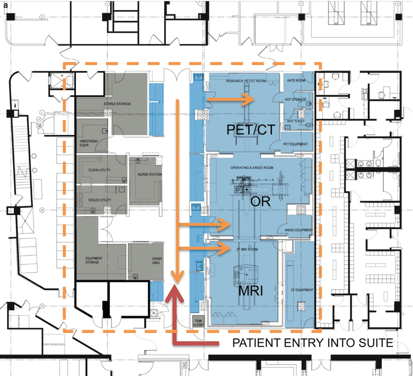
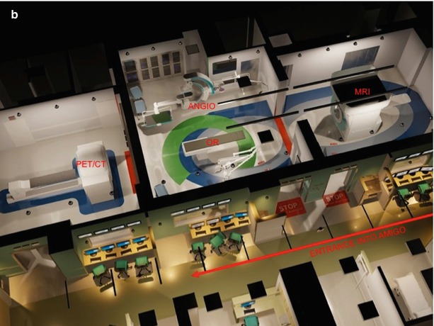
Fig. 24.2
Suite floor plan. (a) Procedure space is to the right of the central corridor and support space is to the left. The red arrow shows the pathway for entry into the suite and the orange arrows show the pathways into the procedure rooms (Courtesy of Payette Architects). (b) Axiometric view of suite (Courtesy of Balazs Lengyel MD)
Structure: AMIGO is advantageously located two floors below grade on the hospital’s subgrade foundation. The larger and stronger reinforced concrete columns on this level support the ceiling-mounted MRI scanner’s structural steel, causing less concern from vibration than placing the suite on an upper floor. The greatest structural challenge was meeting the steel beam deflection tolerance specification that IMRIS required to support the weight of the MRI as it transits: no more than 1/8″ deflection of steel beam for every 8 ft of beam length. The specific challenge was to maintain this requirement while limiting the dimensions of the steel beam in order to allow infrastructure to fit overhead. The unique design, shown in Fig. 24.3, kept the steel support simple and allowed for maximum flexibility for infrastructure to fit within the ceiling plenum.
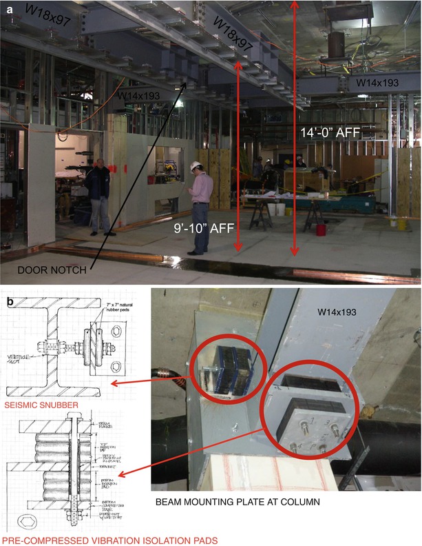
Fig. 24.3
(a) Construction photograph of the steel structure (Courtesy of Payette Architects). (b) Design and construction photograph of the structural isolation damper (Courtesy of Payette Architects & Cavanaugh Tocci Associates)
Another concern was vibration from outside the hospital that might impact the image quality from the MRI. A major source of vibration is the plant that provides supplemental electricity to the six hospitals in the surrounding Longwood Medical area, located adjacent to the end of the hospital housing AMIGO. To mitigate the effects, large pre-compressed vibration isolation pads were designed for all beam and column connections (Fig. 24.3).
Shielding and Penetration: Along with the shielding vendor (ETS-Lindgren, Glendale Height, IL) and IMRIS, the team designed the shielding efficiently to enclose the two impacted rooms. Both the OR and MRI rooms are six-sided copper RF-shielded boxes to prevent electromagnetic interference from impacting MR image quality. Both the OR and PET/CT rooms are lead shielded. The sliding doors and control room doors in the OR were designed with additional structural reinforcement to support the weight of the copper and lead.
The viewing windows into the OR, MRI, and PET/CT rooms (Fig. 24.4) are oversized and comprised of polarizing privacy glass, lead glass, and RF copper mesh glass. The privacy glass when turned off turns opaque and meets the laser safety requirements to allow laser operation inside the room without additional laser safety measures being installed.

Fig. 24.4
PET/CT and OR control room (Courtesy of Warren Jagger Photography)
Silicon rolled steel for magnetic shielding was located only on the rear of the MRI room to prevent the MRI scanner’s fringe field from extending into the hallway adjoining the EP labs, to protect patients, personnel, and equipment outside the suite.
Another unique detail in AMIGO is the inclusion of an independent RF-shielded equipment cabinet inside the larger RF-shielded enclosure. This feature allows clinicians and researchers to stage non-MRI-compatible equipment into the interventional environment via waveguides and filtered connectors without affecting image quality (Fig. 24.5). Additional pen panels were added under each control window as well as the MRI equipment room to allow for additional connections with equipment placed outside the RF rooms. All subpanels are modular and can be retrofitted to meet future user requirements (Fig. 24.5).
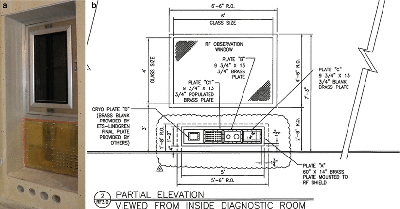
Fig. 24.5
(a) RF-shielded cabinet inside MRI room and penetration panels and waveguides (Courtesy of Payette Architects). (b) Drawing detail of shielded window and under counter penetration panel (Courtesy of Payette Architects)
Location of all filters and waveguides required for all infrastructure (electrical conduits, HVAC ducts, med gas piping, etc.) penetrating the shielded rooms was carefully coordinated to ensure a clean plenum space above the ceiling. Boom mounts were designed to obviate the requirement for kicker supports, a space saving method that allowed for much needed above ceiling space (Fig. 24.6).
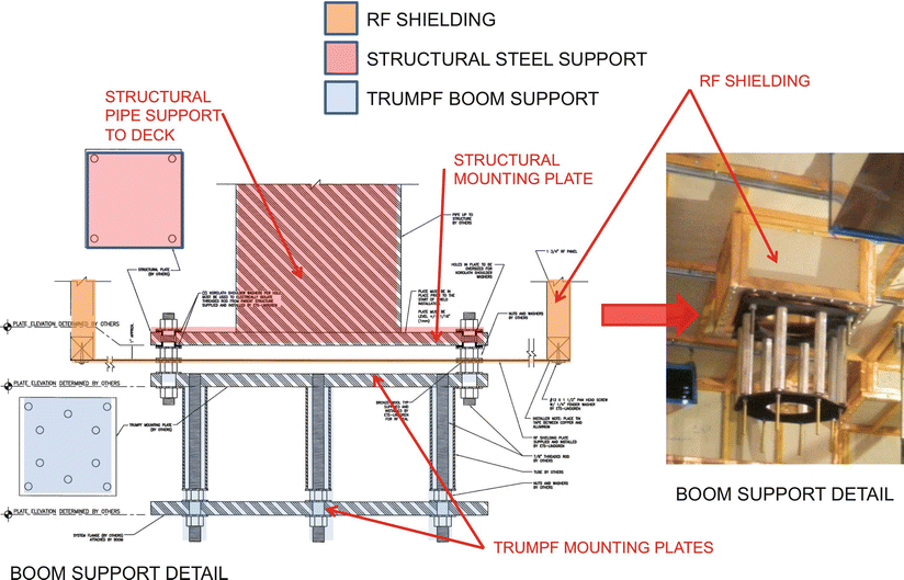
Fig. 24.6


Design detail and photograph of the dielectric isolation and shielding scheme for the boom mounts (Courtesy of Payette Architects & ETS-Lindgren Shielding)
Related posts:


Stay updated, free articles. Join our Telegram channel
Full access? Get Clinical Tree
