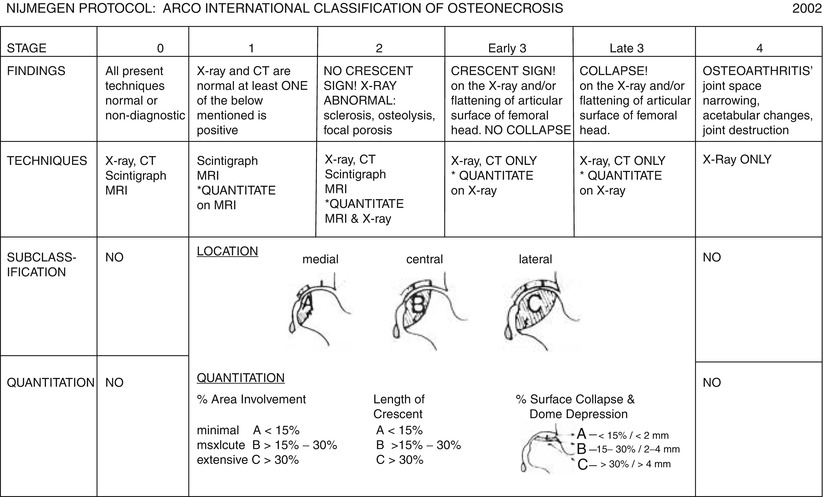
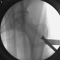
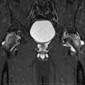
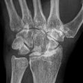
Fig. 28.1
ARCO international classification of osteonecrosis
Stage 0: All diagnostic studies are normal and diagnosis is made by histology only. A painful hip in a patient who has either one or more risk factors for osteonecrosis or who has a proven osteonecrosis on the contralateral side.
Stage 1: A band lesion of a low signal intensity around the necrotic area is seen on MRI scans. No changes are seen on plain radiographs.
Stage 2: Subtle signs of osteosclerosis, focal osteoporosis, or cystic change can be identified in the femoral head on plain radiographs. Still there is no evidence of subchondral fracture.
Stage 3: Early fracture in the subchondral portion is seen on plain radiography or by computed tomography or tomograms.
Stage 4: There is osteoarthritis of the joint with accompanying joint space narrowing, acetabular changes, and finally destruction of the joint.
28.4 Latest Modification in Nijmegen (Fig. 28.2)
Subsequently after using this classification, there was a need to subdivide stage 3 into early and late stages. Sometimes, osteonecrosis in early collapse does not progress to further stage, and patients are doing well for a prolonged time without any intervention. The results of the joint preserving procedures such as bone grafting and osteotomies showed a clear difference between the head with and without collapse. The results in early stage 3 without definite collapse were significantly better than those in late stage 3 with definite collapse. This subdivision seems to be of crucial importance, particularly for the evaluation of outcome after treatment of osteonecrosis. Besides, other classification systems have a separation between the “early” stage before a collapse of the femoral head develops and the “late” stage when a definite collapse is already present.
