(1)
Department of Nuclear Medicine, Kuwait University, Safat, Kuwait
2.1 Introduction
2.2.1 Plasma Membrane
2.2.3 Cytoskeleton
2.2.4 Nucleus
2.3.3 Genetic Code
2.4.1 The Cell Cycle
2.4.2 Mitosis and Cytokinesis
2.4.3 Rates of Cell Division
2.4.4 Chromosomes and Diseases
2.6.1 Normal Growth
2.6.2 Neoplastic Growth
2.7.1 Role of ATP
2.7.2 Production of ATP
2.9.1 Imaging Cell Death
Abstract
The human body contains trillions of cells that are generated by repeated division from a single precursor cell. They constitute clones. With proliferation, some of the cells become differentiated from others, adopting a different structure, different chemistry, and different function.
2.1 Introduction
The human body contains trillions of cells that are generated by repeated division from a single precursor cell. They constitute clones. With proliferation, some of the cells become differentiated from others, adopting a different structure, different chemistry, and different function.
In the human body, more than 200 distinct cell types are assembled into a variety of tissue types such as epithelia, connective tissue, muscle, and nervous tissue. Each organ in the body is an aggregate of many different cells held together by intercellular supporting structures. Although these cells often differ markedly, they all have basic characteristics that are alike.
2.2 Cell Structure and Function
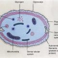
The different substances that make up the cell are collectively called protoplasm, which is composed mainly of water, electrolytes, proteins, lipids, and carbohydrates. The two major parts of the cell are the nucleus and cytoplasm. The cytoplasm is separated from the extracellular fluid by a cell membrane, while the nucleus is separated from the cytoplasm by a nuclear membrane (Fig. 2.1). The major organelles in the cell are organelles derived from membranes, organelles involved in gene expression, and organelles involved in energy production [1]. The important subcellular structures of the cell and their functions are summarized in Table 2.1.
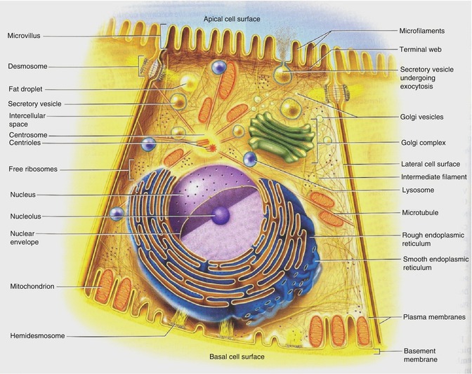
Fig. 2.1
Schematic drawing of the cell clearly depicting the intricate network of interconnecting intracellular membrane structures such as the endoplasmic reticulum (rough and smooth), mitochondria, lysosomes, and nucleus (Reprinted with permission from Saladin [1])
Cell structure | Major functions |
|---|---|
Plasma membrane | Cell morphology and movement, transport of ions and molecules, cell-to-cell recognition, cell surface receptors |
Endoplasmic reticulum | Formation of compartments and vesicles, membrane synthesis, synthesis of proteins and lipids, detoxification reactions |
Lysosomes | Digestion of worn-out mitochondria and cell debris, hydrolysis of proteins, carbohydrates, lipids, nucleic acids |
Peroxisomes | Oxidative reactions involving molecular oxygen, utilization of hydrogen peroxide (H2O2) |
Golgi complex | Modification and sorting of proteins for incorporation into organelles and for export; formation of secretory vesicles |
Microbodies | Isolation of particular chemical activities from the rest of the cell body |
Mitochondria | Cellular respiration; oxidation of carbohydrates, proteins and lipids; urea and heme synthesis |
Nucleus | DNA synthesis and repair; RNA synthesis and control; center of the cell; directs protein synthesis and reproduction |
Chromosomes | Contain hereditary information in the form of genes |
Nucleolus | RNA processing, assembles ribosomes |
Ribosomes | Sites of protein synthesis in cytoplasm |
Cytoplasm | Metabolism of carbohydrates, lipids, amino acids, nucleotides |
Cytoskeleton | Structural support, cell movement, cell morphology |
2.2.1 Plasma Membrane
The plasma membrane encloses the cell, defines its boundaries, and maintains the essential difference between the cytoplasm and the extracellular environment. The cell membranes are assembled from four major components: a lipid bilayer, membrane proteins, sugar residues, and a network of supporting fibers.
The lipid bilayer is a major barrier, impermeable to water-soluble molecules such as ions, glucose, and urea. The major classes of membrane lipid molecules are phospholipids, cholesterol, and glycolipids. The membrane proteins are responsible for most membrane functions such as transport, cell identity, and cell adhesion and constitute transport channels, transport molecules, specific receptors, and enzymes. The cell surface often has a loose carbohydrate coat called glycocalyx. The sugar residues generally occur in combination with proteins (glycoproteins, proteoglycans) or lipids (glycolipids).
2.2.2 Cytoplasm and Its Organelles
Cytoplasm is an aqueous solution (cytosol) that fills the cytoplasmic matrix, the space between the nuclear envelope and the cell membrane. The cytosol contains many dissolved proteins, electrolytes, glucose, certain lipid compounds, and thousands of enzymes. In addition, glycogen granules, neutral fat globules, ribosomes, and secretory granules are dispersed throughout the cytosol. Many chemical reactions of metabolism occur in the cytosol, where substrates and cofactors interact with various enzymes. The various organelles suspended in the cytosol are either surrounded by membranes (nucleus, mitochondria, and lysosomes) or derived from membranous structures (endoplasmic reticulum, Golgi apparatus). All biological membranes are phospholipid bilayers with embedded proteins.
2.2.2.1 The Endoplasmic Reticulum
An interconnecting network of tubular and flat membranous vesicular structures is called the endoplasmic reticulum (ER). Like the cell membrane, the walls of the ER are composed of a lipid bilayer containing many proteins and enzymes. The regions of ER rich in ribosomes are termed rough or granular ER, while the regions of ER with relatively few ribosomes are called smooth or agranular ER. Ribosomes are large molecular aggregates of protein and ribonucleic acid (RNA) that are involved in the manufacture of various proteins by translating messenger RNA (mRNA) copies of genes.
2.2.2.2 The Golgi Complex
The Golgi complex or apparatus is a network of flattened smooth membranes and vesicles. It is the delivery system of the cell. It collects, packages, modifies, and distributes molecules within the cell or secretes the molecules to the external environment. Within the Golgi bodies, the proteins and lipids synthesized by the ER are converted to glycoproteins and glycolipids and collected in membranous folds or vesicles called cisternae, which subsequently move to various locations within the cell. In a highly secretory cell, the vesicles diffuse to the cell membrane and then fuse with it and empty their contents to the exterior by a mechanism called exocytosis. The Golgi apparatus is also involved in the formation of intracellular organelles such as lysosomes and peroxisomes.
2.2.2.3 Lysosomes
Lysosomes are small vesicles formed by the Golgi complex and have a single limiting membrane. Lysosomes maintain an acidic matrix (pH 5 and below) and contain a group of glycoprotein digestive enzymes (hydrolases) that catalyze the rapid breakdown of proteins, nucleic acids, lipids, and carbohydrates into small basic building molecules. In white blood cells, lysosome contents are released into the vacuole around the bacteria and serve to kill and digest those bacteria. Lysosomes also release hydrolytic enzymes into the cytoplasm to digest the entire cell. This is termed programmed cell death (apoptosis) or selective cell death, which is one of the principal mechanisms involved in the removal of unwanted cells and tissues in the body.
2.2.2.4 Peroxisomes
Peroxisomes are small membrane-bound vesicles or microbodies derived from the ER or Golgi apparatus. Many of the enzymes within the peroxisomes are oxidative enzymes that generate or utilize hydrogen peroxide (H2O2). Peroxisomes protect the cell from its own production of toxic hydrogen peroxide. White blood cells, for example, produce hydrogen peroxide to kill bacteria. The oxidative enzymes in peroxisomes break down the hydrogen peroxide into water and oxygen. Peroxisomes are also involved in the oxidative metabolism of long-chain fatty acids.
2.2.2.5 Mitochondria
Mitochondria are tubular or sausage-shaped organelles (1–3 μm). They are composed mainly of two lipid bilayer-protein membranes. The outer membrane is smooth and derived from the ER. The inner membrane contains many infoldings or shelves called cristae which partition the mitochondrion into an inner matrix called mitosol and an outer compartment. The outer membrane is relatively permeable but the inner membrane is highly selective and contains different transporters. The inner membrane contains various proteins and enzymes necessary for oxidative metabolism, while the matrix contains dissolved enzymes necessary to extract energy from nutrients. The total number of mitochondria per cell depends on the specific energy requirements of the cell and may vary from less than a hundred to up to several thousand. Mitochondria are the “powerhouses” of the cell. The cell derives energy from glucose, amino acids, and fatty acids. In a process called glycolysis, glucose is converted to pyruvic acid, which subsequently enters mitochondria where it begins a sequence of chemical reactions called the citric acid or Krebs cycle. Various enzymes present in the inner membrane oxidize the pyruvic acid to carbon dioxide and water. The oxidative metabolism of the glucose molecule generates 36 molecules of ATP. The amino acids and fatty acids are converted to acetyl coenzyme A (in the cytoplasm) which also enters the citric acid cycle and gets oxidized with the generation of ATP molecules.
2.2.2.6 Ribosomes
Ribosomes are large complexes of RNA and protein molecules and are normally attached to the outer surfaces of the ER. The major function of ribosomes is to synthesize proteins. Each ribosome is composed of one large and one small subunit with a mass of several million daltons.
2.2.3 Cytoskeleton
The cytoplasm contains a network of protein fibers, called the cytoskeleton, that provides a shape to the cell and anchors various organelles suspended in the cytosol. The fibers of the cytoskeleton are made up of different proteins of different sizes and shapes such as actin (actin filaments), tubulin (microtubules), and vimentin and keratin (intermediate filaments).
2.2.4 Nucleus
The nucleus is the largest membrane-bound organelle in the cell, occupying about 10 % of the total cell volume. The nucleus is composed of a double membrane, called the nuclear envelope, that encloses the fluid-filled interior, called nucleoplasm. The outer membrane is contiguous with the ER. The nuclear envelope has numerous nuclear pores, permitting certain molecules to pass into and out of the nucleus. The primary functions of the nucleus are the control of cell division and the phenotypic expression of genetic information that directs all of the activities of a living cell. The cellular deoxyribonucleic acid (DNA) is located in the nucleus as a DNA-histone protein complex known as chromatin that is organized into chromosomes. The total genetic information stored in the chromosomes of an organism is said to constitute its genome. The human genome consists of 24 chromosomes (22 different chromosomes and two sex chromosomes). The smallest unit of DNA that encodes a protein product is called a gene and consists of an ordered sequence of nucleotides located in a particular position on a particular chromosome. There are approximately 100,000 genes per human genome, and only a small fraction (15 %) of the genome is actively expressed in any specific cell type. The genetic information is transcribed into ribonucleic acid (RNA), which subsequently is translated into a specific protein on the ribosome. The nucleus contains a subcompartment called the nucleolus that contains large amounts of RNA and protein. The main function of the nucleolus is to form granular subunits of ribosomes, which are transported into the cytoplasm where they play an essential role in the formation of cellular proteins.
2.3 The Genetic Material and Gene Expression
2.3.1 The Genetic Material: DNA
The ability of cells to maintain a high degree of order depends on the hereditary or genetic information that is stored in the DNA [5–9]. Within the nucleus of all mammalian cells, a full complement of genetic information is stored, and the entire DNA is packaged into chromosomes.
2.3.1.1 DNA Structure
DNA was first discovered in 1869 as a white substance from the cell nuclei of human pus and was called “nuclein.” Since nuclein was slightly acidic, it was known as nucleic acid. In the 1920s, two sorts of nucleic acids (DNA and RNA) were identified. The structure of DNA molecule as a polynucleotide was later shown to be formed by the polymerization of nucleotides. Each nucleotide subunit of DNA molecule is composed of three basic elements: a phosphate group, a five-carbon sugar (deoxyribose), and one of the four types of nitrogen-containing organic bases. Two of the bases, thymine and cytosine, are called pyrimidines, while the other two, adenine and guanine, are called purines.
Although some forms of cellular DNA exist as single-stranded structures, the most widespread DNA structure represents DNA as a double helix containing two polynucleotide strands that are mirror images of each other (Fig. 2.2). The “backbone” of the DNA molecule is composed of the deoxyribose sugars joined by phosphodiester bonds to a phosphate group, while the bases are linked in the middle of the molecule by hydrogen bonds. The relationship between the bases in a double helix is described as complementarity, since adenine always bonds with thymine and guanine always bonds with cytosine. As a consequence, the double-stranded DNA contains equal amounts of purines and pyrimidines.
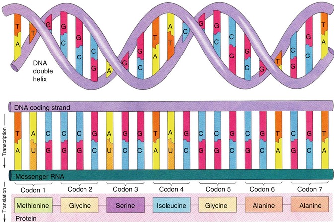
Fig. 2.2
The double-stranded DNA molecule consists of bases, deoxyribose sugar, and phosphate. Note the hydrogen bonds between the two strands of DNA molecules (Reprinted with permission from Saladin [1])
2.3.1.2 DNA Replication
All the chromosomes in the nucleus duplicate their DNA prior to every cell division in order to serve as genetic material. When a DNA molecule replicates, the double-stranded DNA separates or unzips at one end, forming a replication fork (Fig. 2.3). The process of replication proceeds by a mechanism in which a new DNA strand is synthesized that matches each of the original strands serving as a template.
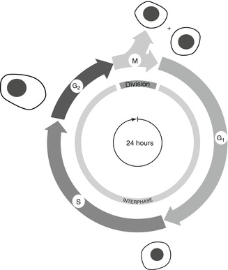
Fig. 2.3
The cell division cycle is generally represented by four successive phases. During the interphase, the cell grows continuously, and only during M phase does it undergo division. DNA replication occurs during S phase, while G1 and G2 are the gaps during which cells normally show additional growth such as protein and enzyme synthesis. Cells in G1, if they are not committed to DNA replication (that is, entering S phase), may enter into a resting state, often called G0, where they can remain for days, or even years, before resuming proliferation (Reprinted with permission from Alberts et al. [13])
At the end of each round of replication, one of the parental strands is maintained intact, and it combines with one newly synthesized complementary strand.
2.3.1.3 DNA Mutation
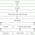
A mutation is any inherited change in the genetic material involving irreversible alterations in the sequence of DNA nucleotides. These mutations may be phenotypically silent (hidden) or expressed (visible). Mutational damage to DNA is generally caused by one of three events: (a) Ionizing radiation causes double-stranded breaks in DNA due to the action of free radicals on phosphodiester bonds. (b) Ultraviolet radiation creates DNA cross-links due to the absorption of UV energy by pyrimidines. (c) Chemical mutagens modify DNA bases and alter base-pairing behavior. Mutations in germ line tissue are of enormous biological significance, while somatic mutations may cause cancer.
2.3.1.4 DNA Recombination
DNA can undergo exchange events through recombination leading to genetic material rearrangement. Recombination is defined as the creation of new gene combinations and may include exchange of an entire chromosome or rearranging the position of a gene or a segment of a gene on a chromosome.
2.3.2 Gene Expression and Protein Synthesis
Heredity is the ability of the cell to use the information in its DNA to control and direct the synthesis of all proteins in the body, as proteins are the tools of heredity. The production of RNA is called transcription and is the first stage of gene expression. The result is the formation of messenger RNA (mRNA) from the base sequence specified by the DNA template. All types of RNA molecules are transcribed from the DNA. The mRNA molecules are finally transported to the ER in the cytoplasm, where proteins are synthesized.
Related posts:
Stay updated, free articles. Join our Telegram channel

Full access? Get Clinical Tree


