The Fetal Circulation
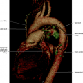
Early in the fetal life, the fetal blood reaches the placenta through the two umbilical arteries and returns to the fetus by the two umbilical veins. Later, the right umbilical vein disappears and the left vein persists as the single returning vessel. The fetal blood receives oxygen and nutrients by close contact with maternal blood in the placenta. The umbilical vein (persistent left umbilical vein) enters the abdomen at the umbilicus and runs along the edge of the falciform ligament to the hepatic visceral surface, where it sends branches to the left hepatic lobe and joins the left branch of the portal vein. At the opposite side of these anastomoses arises the ductus venosus, which joins the inferior vena cava, conveying the oxygen-rich blood that comes from the maternal placenta. The fetal portal vein is small, and the right and left branches function as branches of the ductus venosus, carrying oxygenated blood to the liver. At the inferior vena cava, the oxygenated blood mixes with a small amount of oxygen-poor blood from the caudal portion of the fetus. The blood from the inferior vena cava together with the blood from the ductus venosus enters the right atrium and hits the interatrial membrane and is directed through the foramen ovale into the left atrium, guided by the valve of the inferior vena cava. At the left atrium the oxygen-rich blood mixes with a small amount of nonoxygenated blood from the pulmonary vein. From the left atrium, the blood enters the left ventricle and, subsequently, the aorta. A small portion of oxygenated blood, instead of crossing the foramen ovale, joins the blood flow from the superior vena cava and, after passing through the right atrium, enters the right ventricle of the heart. The inflow from the superior vena cava plus the small amount of blood from the umbilical vein is diverted to the pulmonary artery, thereby supplying the lungs. Most of this blood flow, however, is shunted through the ductus arteriosus directly into the descending aorta, where it joins the stream of blood ejected from the left ventricle. Most of the oxygenated blood ejected from the left ventricle reaches the heart and brain circulation, providing higher oxygen content to these organs rather than to structures less sensitive to hypoxia in the abdomen and extremities. The blood in the descending aorta is poorer in oxygen and is partly distributed to the lower limbs and viscera of the abdomen and pelvis, but most of it returns to the placenta via the umbilical arteries, branches of the internal iliac arteries (Fig. 1.1).
Stay updated, free articles. Join our Telegram channel

Full access? Get Clinical Tree


