The Lower Urinary Tract
Cathie Scholl
|

OBJECTIVES
Describe the embryologic development, normal anatomy, and function of the lower urinary tract.
Discuss the various sonographic techniques that can be used for evaluation of the lower urinary tract.
Identify the normal sonographic appearance of the lower urinary tract and common anatomic variants.
List clinical indications associated with lower urinary tract disease.
Describe the sonographic appearance of congenital lower urinary tract abnormalities such as exstrophy, duplication, posterior urethral valves, ectopic ureter, and ureterocele.
List the common causes and sonographic appearance of cystitis.
Identify the sonographic appearance of reflux, neurogenic bladder, and bladder wall abnormalities.
Describe common mechanisms for bladder trauma and the appearance of pathologies related to trauma.
List common causes of bladder wall thickening.
Describe the sonographic appearance of benign and malignant bladder tumors.
Define stress incontinence and describe sonographic techniques used in the diagnosis.
Identify technically satisfactory and unsatisfactory sonographic examinations of the lower urinary tract.
KEY TERMS
bladder flap hematoma
cystitis
diverticula
ectopic ureter
exstrophy
neurogenic bladder
posterior urethral valves
squamous cell carcinoma
stress incontinence
transitional cell carcinoma
urachal cyst
ureterocele
vesicoureteral reflux
GLOSSARY
cystoscopy
procedure in which a scope is used to evaluate the urethra, bladder, and pelvic ureters
procedure in which a scope is used to evaluate the urethra, bladder, and pelvic ureters
hematuria
presence of red blood cells in the urine; hematuria can be microscopic (not visible with the naked eye) or macroscopic
presence of red blood cells in the urine; hematuria can be microscopic (not visible with the naked eye) or macroscopic
trabeculated bladder
thickened, irregular bladder wall frequently seen in patients with long-standing obstruction or neurogenic bladder
thickened, irregular bladder wall frequently seen in patients with long-standing obstruction or neurogenic bladder
voiding cystourethrogram (VCUG)
a procedure used to evaluate for urinary reflux in which the patient is catheterized and the bladder is filled with a contrast agent; the bladder is examined under fluoroscopy to evaluate for vesicoureteral reflux both before and during patient voiding
a procedure used to evaluate for urinary reflux in which the patient is catheterized and the bladder is filled with a contrast agent; the bladder is examined under fluoroscopy to evaluate for vesicoureteral reflux both before and during patient voiding
The lower urinary tract consists of the pelvic ureters, bladder, and urethra. Whenever a reference is made to the urinary system, the kidneys come to mind first. The ureter, bladder, and urethra are also part of the urinary system and play important roles in transporting, storing, and eliminating urine. The pelvic ureter and urethra are conduits in the process of elimination of urine. The bladder is located anatomically between these two structures and functions as a reservoir for urine storage. The primary focus of this chapter is the urinary bladder. The normal pelvic ureter and the urethra are not usually seen sonographically, but these structures may be visualized with coexisting pathologic conditions.
The urine-filled bladder is one of the most accessible abdominopelvic organs for sonography examinations. Recognition of normal bladder anatomy, including its position, size, shape, and appearance, helps the sonographer identify congenital anomalies of the bladder, pathologies, and abnormalities in the surrounding anatomy.
ANATOMY AND ORGANOGENESIS
During early embryology of the human urogenital system, three sets of kidneys develop—the pronephros (early in the 4th embryologic week), mesonephros (late in the 4th week), and the metanephros (5th week)—in three successive waves, from cranial to caudal, with the third, most inferior pair of kidneys (metanephros) becoming the permanent kidneys.1 The caudal end of the hindgut has a dilated chamber, the cloaca. The cloacal endoderm is in close contact with the surface ectoderm, and together, they form the cloacal membrane. An extension from the cloaca into the umbilical cord is the allantois. The intermediate mesoderm of the gastrula bulges into the dorsal aspect of the intraembryonic coelom as a urogenital ridge on each side. This further develops into two ridges: a medial genital (gonadal) ridge and a lateral nephrogenic ridge (or cord). A mesonephric (wolffian) duct and paramesonephric (müllerian) duct form in the nephrogenic ridge or cord.1
In approximately the 7th gestational week, the urorectal septum between the allantois and hindgut fuses with the cloacal membrane, dividing it into the ventral (anterior) urogenital sinus and a dorsal (posterior) rectum.2 The upper part of the urogenital sinus is the fusiform bladder. The lower pelvic and phallic parts of the urogenital sinus form the urethra and related glands and structures in each sex. The wolffian ducts give rise to the ureters; they also form the efferent tubules, duct of the epididymis, vas deferens, seminal vesicles, and ejaculatory ducts in males and the epoophoron, paroophoron, and Gartner duct in females.
The ends of the mesonephric ducts (wolffian and müllerian) and the endodermal cloaca form the urinary bladder.1 The cloaca—the terminal, caudal, blind-ended portion of the hindgut—is the major structure that forms the lower part of the urinary and genital tract. Its primary function is to serve as the primitive receptacle into which the reproductive and excretory tracts empty.
The metanephric duct (future ureter) develops from a ureteric bud growing from the caudal end of the mesonephric duct. In a short time, the metanephric duct shifts anteriorly and makes its own connection with the cloaca/urogenital sinus/bladder.
At the 8th week of gestation, all embryos have identical primordia in the indifferent stage of urogenital development, with gonads capable of developing into testes or ovaries. In males, the paramesonephric (müllerian) ducts degenerate. The mesonephric ducts become the ductus deferens, ejaculatory ducts, and seminal vesicles. The urogenital sinus develops into the urinary bladder, prostate gland, bulbourethral glands (Cowper gland), paraurethral glands, and prostatic, membranous, and penile (spongy) urethra.1 In females, the mesonephric (wolffian) ducts degenerate, and the paramesonephric ducts develop into the uterine tubes, uterus, and upper part of the vagina. The urogenital sinus forms the bladder, urethra, greater vestibular and paraurethral glands, vestibule, and lower part of the vagina.1
Initially, the bladder is contiguous with the allantois, which eventually becomes a fibrous cord, the urachus (known as the median umbilical ligament in the adult).3 The urachus extends from the apex of the bladder to the umbilicus. In infants and children, the urinary bladder is an abdominal organ until after puberty when it becomes a true pelvic structure.3,4
Urinary Bladder
The bladder develops into a hollow, smooth, musculomembranous, collapsible sac that acts as a reservoir for urine. The bladder is in the retroperitoneum on the pelvic floor just posterior to the pubic symphysis.5 Its size, position, and relationship to other organs vary according to the amount of fluid it contains.2 The urinary bladder is lined with a mucous membrane of transitional epithelium that allows for expansion. This mucous membrane lining contains rugae or folds. When the bladder is empty, the membrane appears folded or wrinkled.5 The mucous membrane is loosely attached to the underlying muscle coat except at the trigone region, where it is firmly attached to the muscular coat, appears smooth, and does not expand during bladder filling.
The bladder is capable of considerable distention because of the lining’s elasticity and rugae and the wall’s elasticity.5 It is uniquely situated in the pelvic cavity for its function of urine storage. Bladder capacity varies greatly and depends on many factors, including the age and physical condition of the patient. The normal adult bladder is generally moderately full at 500 mL (a pint) of urine, but it may hold nearly double that if necessary.3,5
Normally, the bladder is a round-edged tetrahedron with a superior, a posterior, and two inferior surfaces. The superior surface has two regions: the fundus, located posteriorly, and the apex, located anteriorly. The two ureteral orifices are located in the body on the posteroinferior portion. The urethral orifice is located in the neck of the bladder and is the most inferior region.1
When the bladder is empty, the anterior surface lies just behind and, rarely, superior to the symphysis in both males and females.1 The fibrous medial umbilical ligament (obliterated urachus) extends from the apex upward as a blunt cone with a solid, slender continuation in the midline of the abdominal wall and attaches to the umbilicus.1
Related anatomy in the pelvis varies depending on the quantity of urine and with the condition of the rectum, being pushed upward and forward when the rectum is distended.1
When distended with urine, the bladder can rise approximately 16 cm above the symphysis pubis. The bladder ascends into the abdominal cavity and meets with the lower anterior abdominal wall. When fully distended, it can be readily palpated or percussed. As the bladder enlarges, it loses its ovoid or spherical configuration and becomes more globular. Coils of the small intestine lie adjacent to the upper surface of the bladder and are displaced posteriorly as the bladder enlarges. With overdistention, such as acute or chronic urinary retention, the lower abdomen may visibly bulge.
When the bladder is relatively empty in the female, the fundal region of the bladder lies in contact with the anterior wall of the vagina and cervix (Fig. 13-1A). The uterus and vagina are interposed between the bladder and the rectum.3 When the bladder is empty, the uterus rests on the bladder’s superior surface. Female reproductive and pelvic muscular anatomy is greatly enhanced using the traditional full-bladder technique.
In the male, the fundus and the body of the bladder are related to the rectum, separated above by the rectovesical pouch of the peritoneum and inferolaterally on each side by a ductus deferens and seminal vesicle.3 The prostate is a fibromuscular and glandular organ that lies just inferior to the bladder.3 The base of the prostate is applied to the caudal surface of the bladder. The greater part of this surface is directly continuous with the bladder wall. The normal prostate encircles the prostatic urethra and prostate gland secretion enters the prostatic urethra via several ducts. The seminal vesicles lie just cephalad to the prostate under the base of the bladder. They are approximately 6 cm long and quite soft. Each vesicle joins its corresponding vas deferens to form the ejaculatory duct (Fig. 13-1B, C).
Trigone
On the floor of the bladder, a triangular region, the trigone, has no rugae and is firmly attached to the muscular coat.6 The trigone is outlined by the three openings in the bladder: two from the ureters and one into the urethra (Figs. 13-1C and 13-2). The ureteral orifices are situated superiorly and laterally at the extremities of the crescent-shaped interureteric ridge that forms the proximal border of the trigone.1 The urethral opening is located at its anterior, midline, lower corner at the bladder neck.
Ureters
The ureters are slender tubes that convey urine from the kidneys to the bladder.6 Each ureter is a continuation of the renal pelvis. From there, they descend in the retroperitoneum and run obliquely through the posterior bladder wall. The average length of the ureter is 30 cm and the diameter is 6 mm.6 The ureters are constricted in three places: (1) at the ureteropelvic junction, (2) as they cross the iliac vessels, and (3) at the junction with the bladder.
The distal ureter enters obliquely through the bladder wall by slit-like openings. This anatomic arrangement prevents the backflow of urine. As the bladder fills, the pressure increases, causing the upper and lower walls of the terminal portions of the ureter to become closely applied to each other, acting as valves to prevent regurgitation of urine from the bladder. When the bladder is distended, the openings of the ureters are about 5 cm apart; however, the distance between them is diminished by half when the bladder is empty and contracted.
Urethra
The urethra is a thin-walled fibromuscular tube that drains urine from the bladder and conveys it outside the body. The urethra represents the terminal portion of the urinary tract. At the bladder-urethral junction, a thickening of the detrusor smooth muscle of the bladder wall forms the internal urethral sphincter. This involuntary muscle keeps the urethra closed and prevents leaking between voiding. The sphincter is unique in that contraction opens it and relaxation closes it. The external urethral sphincter surrounds the urethra as it passes through the urogenital diaphragm. This sphincter is formed of skeletal muscle and is controlled voluntarily.5,6
The length and functions of the urethra differ in males and females. The female urethra is 3 to 4 cm long and functions only to convey urine from the body.5 It lies directly posterior to the symphysis pubis and anterior to the vagina. The external urethral orifice—the external opening of the urethra—lies anterior to the vaginal opening and posterior to the clitoris. The opening of the urethra to the exterior is referred to as the urinary meatus.
The male urethra serves a double function: a conduit for eliminating urine and also as the terminal portion of the reproductive system serving as the passage for ejaculate (semen). The male urethra is approximately 20 cm long and has three regions. The prostatic urethra, about 2.5 cm long, runs within the prostate.5 The membranous urethra, which runs through the urogenital sinus, extends about 2 cm from the prostate to the beginning of the penis. The spongy urethra passes through the penis and opens at its tip—the external urethral orifice.
PHYSIOLOGY
The mechanism for voiding urine (micturition) starts with involuntary and voluntary nerve impulses.5 Even though the bladder has a greater capacity, when the volume of urine exceeds 200 to 400 mL, stretch receptors trigger transmission of impulses to the lower portion of the spinal cord, initiating the conscious desire to expel urine and a subconscious reflex, the micturition reflex.7 The combination of voluntary relaxation of the external sphincter muscle of the bladder, reflex contraction of linear smooth muscle fibers along the urethra, and then contraction of the detrusor muscle squeezes urine out of the bladder.5 Parasympathetic fibers transmit the impulses that cause contractions of the bladder and relaxation of the internal sphincter.5,7,8
Because the external sphincter is under voluntary control, we can choose to postpone bladder emptying. Voluntary contraction of the external sphincter to prevent or terminate micturition is learned and is possible only if the nerves supplying the bladder and urethra—the projection tracts of the cord and brain—and the motor area of the cerebrum are all intact.7,8 Incontinence—involuntary emptying of the bladder—results from aging or trauma to any of these parts of the nervous system by cerebral hemorrhage or cord injury.5, 6 and 7
Retention is an inability to empty the bladder even though the bladder contains an excessive amount of urine.9 Catheterization may be used to relieve the discomfort accompanying
retention. 30% of patients who are catheterized routinely eventually develop a “ledge” posteriorly at the bladder neck from catheter trauma. The ledge makes voiding difficult and considerably complicates the catheterization process.
retention. 30% of patients who are catheterized routinely eventually develop a “ledge” posteriorly at the bladder neck from catheter trauma. The ledge makes voiding difficult and considerably complicates the catheterization process.
 FIGURE 13-1 Normal anatomy. Coronal planes of the (A) female and (B) male pelvis show the normal anatomic relationship of the bladder with surrounding structures. |
 FIGURE 13-1 (continued) C: This illustration shows the interior cutaway sections of the male urinary bladder and the prostatic urethra and a topographic anatomy of the male pelvic organs. |
 FIGURE 13-2 Trigone. Three-dimensional rendered image of the bladder trigone demonstrating ureteral and urethral openings (arrows). The three-dimensional rendered image of the inner bladder lining provides a virtual cystogram of the bladder wall. (Image courtesy of Philips Medical Systems, Bothell, WA.) |
SONOGRAPHIC EXAMINATION TECHNIQUE
Patient Preparation
To visualize the bladder with the transabdominal approach, it is important that the patient prepares properly. Bladder distention is essential for optimal visualization of the bladder, bladder wall, and related anatomy. Filling the bladder can be accomplished by three methods: (1) instructing the patient to drink 16 oz of water 1 hour before the examination and not to void until the examination is completed; (2) instructing the patient not to void before the examination; or (3) catheterizing the patient and instilling fluid into the bladder through a Foley catheter. Foley catheters are not inserted routinely to fill a bladder unless it is a medical emergency. There have been many studies showing that catheter insertion may introduce infectious contaminants into the body. A Foley catheter balloon will appear as a round cystic structure in the filled bladder and may cast shadows in areas of interest.
A fully distended bladder serves as a cystic reference in the abdominopelvic anatomy, pushes adjacent bowel and gas out of the field of view, and provides a sonographic “window” to identify pelvic anatomy. Routinely in males, the bladder, seminal vesicles, prostate, and rectum are imaged. Routinely in females, the vagina, bladder, uterus, ovaries, adnexa, and rectum are imaged. A full bladder also facilitates identification of dilated ureters. It is not necessary to restrict the diet or use catheters or enemas to reduce intestinal contents or air. Disease processes in pelvic structures can involve or mimic those of other closely related anatomy. Knowledge of pelvic anatomy, including the genitourinary tract, gastrointestinal tract, and pelvic vasculature and musculature is important.
A suitable coupling agent such as ultrasonic gel is used on the skin surface. Transducer selection should take into consideration body habitus and examination objectives. The highest-frequency transducer possible should be selected for scanning, making sure that penetration is adequate to visualize the posterior aspect of the areas of interest.
Scanning Techniques
The most widely used approach to scan the urinary bladder is the transabdominal method.3 The patient is usually examined in the supine position. Sometimes, it is necessary to position the patient obliquely or to roll the patient into a lateral decubitus position to better demonstrate bladder wall abnormalities, the movement of debris or stones to the dependent bladder wall, or bladder tumors. The endovaginal, endorectal, and transperineal methods may also be used to a lesser extent.
The lower urinary tract should be scanned in both longitudinal and transverse planes transabdominally and may be scanned in longitudinal and coronal planes endovaginally and transperineally.3 Using the endorectal approach, the proximal urethra can be visualized and the distal urethra is identified during penile artery evaluation.4 Recent studies have also used translabial and transperineal approaches to evaluate female urethra and urogenital disorders such as stress incontinence.
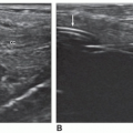
The normal distended urinary bladder appears as an anechoic structure with visible demarcation of the echogenic smooth bladder walls.3 The bladder wall is seen as a smooth echogenic interface and should be of uniform thickness. The thickness of the bladder wall will vary from less than 3 mm when fully distended to 5 mm when nearly empty.3,4,10,11 A pathologically thickened bladder wall is better visualized when the bladder is fully distended.4 If larger than 6 mm when empty or partially distended, the bladder wall should be interrogated for a pathologic process. When the bladder is scanned transabdominally, reverberation echoes are often seen anteriorly in the near field of the bladder image.4,11 Many times, artifacts such as reverberations, grating lobes, and shadowing can be eliminated by sonography system controls and/or altering the transducer position to change the angle of the transmitted sound wave to avoid refraction from abdominal musculature4 (Fig. 13-3A). Equipment manufacturers have added harmonics, speckle reduction, spatial compounding, and other computerized techniques
to aid in the elimination of artifact echoes. Although the normal ureters and urethra are not routinely visualized on transabdominal sonography, their location should be examined because they can be identified in some anomalies, diseases, and pathologic processes.
to aid in the elimination of artifact echoes. Although the normal ureters and urethra are not routinely visualized on transabdominal sonography, their location should be examined because they can be identified in some anomalies, diseases, and pathologic processes.
 FIGURE 13-3 Transverse plane of normal anatomy. A: Reverberation artifact echoes are seen in the near field of a urine-filled bladder (arrow), and the vaginal canal is identified posterior to the bladder. B: The urethral orifice can be identified exiting the trigone (arrow). C: The ureteral orifices are seen as small mucosal elevations (arrows) as they enter the bladder. D and E: Longitudinal plane of normal anatomy. D: On the left, near the midline, the left ureteral orifice is seen entering the trigone. E: The right ureteral orifice is seen entering the trigone. BL, urinary bladder; OV, ovary; UT, uterus. (E: Image courtesy of Steven D. Hatch, Logan, UT.) |
In females, the pelvic segment of the ureters and urethra can be evaluated with transvaginal sonography. The distal ureter can be identified by imaging in a longitudinal scan plane and moving laterally to the pelvic side wall.12 The ureters appear as long tubular hypoechoic structures extending from the lateral aspect of the bladder base to the common iliac vessels.13
If the ureters are not readily seen in the sagittal scan plane, the transverse plane should be used to identify the urethral orifice. Then the probe should be slowly moved upward (anterior) to visualize the ureteral orifices near the lateral portion of the trigone.13 Once the orifices have been identified, the transducer should be rotated to the longitudinal scan plane to lengthen out the distal ureters.
Owing to the proximity of the pelvic segments of the ureters to the common iliac vessels, color Doppler imaging may need to be used to differentiate the structures. When color Doppler imaging is applied, a ureter will not demonstrate color flow, whereas normal vessels should fill with color.
The sonographer should identify the predictable contours of the urine-filled bladder and the smooth echogenic bladder wall. If the patient has never had bladder or pelvic surgery, any deviation from the normal bladder shape, especially asymmetry, should be considered abnormal and a thorough investigation should be performed of the site of the distortion to rule out a mass. On transverse sections, the bladder should appear symmetric. Superiorly, the bladder appears rounded, but in scanning more inferiorly, it appears square owing to the parallel walls of the acetabulum (Fig.13-3B, C). On longitudinal sections, the bladder appears almost triangular, with the base of the triangle parallel with the anterior abdominal wall. In both longitudinal and transverse scans, the lateral walls appear straight or slightly indented by prominent iliopsoas muscles (Fig. 13-3D, E). As different pelvic structures are encountered, it may be necessary to angle the transducer caudad and cephalad and medial to lateral. The symphysis pubis acts as a point of reference on the body surface, where the transducer can be rocked superiorly and inferiorly on longitudinal scans to better view the superior and inferior aspects of the bladder. Transverse imaging must include tilting or a cross-plane imaging by angling the transducer from side to side while continuing to use the fluid in the bladder as a window. This produces a sharper image of the bladder wall and gives a complete sweep to interrogate the entire bladder. Longitudinal and transverse images are more easily interpreted if they are in a sequential order: right to left in the longitudinal plane with the midline image identified and inferior to superior in the transverse plane. Correct labeling must appear on all sonographic images, indicating the location of the scan, patient position, and scanning plane.
Frequently, it is possible to visualize the prostate and seminal vesicles in males using the transabdominal approach. When the transducer is angled caudad under the symphysis pubis, the prostate is seen posteroinferior to the bladder. On a longitudinal scan, the prostate appears as a heterogeneous structure at the most inferior aspect of the bladder. On a transverse image, it appears rounded. Although newer transducers with better penetration and resolution enable sonographers to identify the prostate transabdominally in the adult male, the best way to visualize the prostate is via the endorectal approach (see Chapter 14). The seminal vesicles are seen as two small, oval, hypoechoic structures posterior to the bladder and superior to the prostate (Fig. 13-4A, B).
In patients who are catheterized, the catheter has an anechoic appearance with an echogenic margin and center. If air was used to secure the catheter’s position, it may cause a shadow artifact. Sonographically, the symmetry of the catheter is identifiable as an echogenic incomplete circular structure in a urine-filled bladder (Fig. 13-5).
Routine bursts of echoes—the ureteral jet phenomenon—are seen entering the bladder from the region of the trigone.1,3 At intervals of 5 to 20 seconds, a jet of low-intensity echoes, which lasts a few seconds, starts at the area of the ureteral orifices and flows toward the center of the bladder. Ureteral jets can occur simultaneously, but more commonly, they are separated (Fig. 13-6). Jets can be individually identified
on longitudinal images, but both may be seen simultaneously on a transverse image. Such jets extend up to 3 cm and broaden. After a few seconds, the low-intensity echoes become distributed in the bladder and lose intensity until they can no longer be distinguished. Color Doppler imaging is more sensitive and aids in demonstrating ureteral jets.3 Although evaluation of ureteral jets with color Doppler imaging it cannot predict reflux, the analysis of ureteral jets with color Doppler imaging has been used successfully to determine the degree of ureteral obstruction with unilateral ureteral calculi, with either no detectable ureteral jets or continuous low-level jets on the symptomatic side.4 In the case of bladder diverticulum, there is evidence of reversed flow of urine through the communication between the diverticulum and bladder when slight pressure is applied to the lower abdomen.
on longitudinal images, but both may be seen simultaneously on a transverse image. Such jets extend up to 3 cm and broaden. After a few seconds, the low-intensity echoes become distributed in the bladder and lose intensity until they can no longer be distinguished. Color Doppler imaging is more sensitive and aids in demonstrating ureteral jets.3 Although evaluation of ureteral jets with color Doppler imaging it cannot predict reflux, the analysis of ureteral jets with color Doppler imaging has been used successfully to determine the degree of ureteral obstruction with unilateral ureteral calculi, with either no detectable ureteral jets or continuous low-level jets on the symptomatic side.4 In the case of bladder diverticulum, there is evidence of reversed flow of urine through the communication between the diverticulum and bladder when slight pressure is applied to the lower abdomen.
 FIGURE 13-4 Normal male anatomy. A: In the transverse scan plane, the prostate is posterior to the bladder. B: In the sagittal scan plane, the prostate is inferior to the bladder. (Images courtesy of Susan R. Stephenson.) |
 FIGURE 13-5 Foley catheter (FC). Longitudinal image of the urinary bladder; a FC can be identified within the bladder lumen. The catheter appears to be anechoic, with an echogenic exterior. Note the thickened bladder wall (arrows). (Image courtesy of Philips Medical Systems, Bothell, WA.) |
 FIGURE 13-6 Transverse planes of normal ureteral jets. A: Simultaneous jets of low-intensity echoes (arrows) are visualized entering the urinary bladder. B: Color Doppler image demonstrates both right and left ureteral jets on the transverse image on a female patient. |
If either of the ureters is dilated, the dilated ureter can be visualized as a round, anechoic structure posterior to the bladder in the transverse plane. In the longitudinal plane, the dilated ureter can be visualized as a long, linear structure, usually posterior and to the right or left of the midline.
Bladder volume can be calculated using the formula for an ellipsoid (transverse × anteroposterior [AP] × length × 0.52).3 The scanning equipment usually has computerized techniques to calculate volumes. The systems will calculate bladder volume once the three measurements have been entered. Bladder capacity should be noted. The capacity decreases in association with large pelvic masses, in urinary and pelvic inflammatory disease, prostatic hypertrophy, in patients receiving radiation therapy, in advanced stages of tumor infiltration, and after recent surgery.
Frequently, a postvoid residual volume calculation is also indicated.3 To document the presence of residual urine and calculate its volume, the patient should be asked to empty the bladder. The longitudinal, AP, and transverse measurements are repeated, and another bladder volume is calculated for comparison. Determining the amount of residual urine in patients with suspected bladder outlet obstruction has improved the treatment of these patients. Residual volume increases with age, atonic bladders, bladder neck obstruction, long-term cystitis, and advanced invasion by cancer.
Related posts:

Stay updated, free articles. Join our Telegram channel

Full access? Get Clinical Tree


