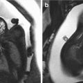
The role of prenatal ultrasound is to diagnose the cleft, establish the extent of the lesion, and diagnose any associated abnormalities, both structural and chromosomal.
4.2 Assessment with 2D ultrasound
The evaluation with 2D ultrasound of the lip and palate is performed using the three spatial planes: sagittal, coronal, and axial or transverse/cross sectional (Fig. 4.2). The profile is obtained by scanning the face in the sagittal plane. The view of the nose and lips is obtained by scanning the face in an anterior coronal section. Finally, the assessment in an axial or cross-sectional plane just below the nose at the level of the upper lip enables visualization of the integrity of the upper lip, as well as the surrounding alveolus, or primary palate.


Fig. 4.2
Evaluation with 2D ultrasound of the lips and palate. (a) Sagittal plane showing the forehead, nose, upper and lower lips, and the chin. (b) Anterior coronal plane showing nasal columella, nostrils, philtrum, upper and lower lips, and chin. (c) Axial plane showing upper lip and primary palate with the alveolar holes. The secondary palate is not seen
The sagittal plane can detect a wide variety of profile alterations during the routine anatomy assessment when certain facial anomalies are present. These include anomalies of the orbits, facial mass, micrognathia, retrognathia, and the absence or hypoplasia of the nasal bones.
The premaxilla, or paranasal protuberance was described by Nyberg et al. [8, 9] in 1992 and 1993. A paranasal mass identified by ultrasound during the second trimester is an important finding of a complete bilateral CLP (Fig. 4.3). In the studies by Nyberg et al. [8, 9], the majority of the fetuses with bilateral CLP demonstrated presence of this mass. The paranasal protuberance can be seen below the nose and represents the prolabium and the premaxilla, composed of the central portion of the upper lip and the primary palate with the alveolar holes of the two central incisors that are derived from the frontonasal processes. The paranasal protuberance can also be evident in cases of larger unilateral CLP.
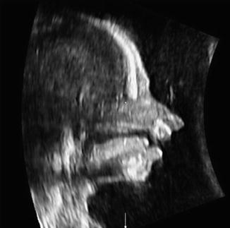

Fig. 4.3
Sagittal plane obtained with 2D ultrasound of a fetus of 22 weeks with a bilateral CLP. A premaxilla or paranasal protuberance is seen, representing the prelabium and the premaxilla projecting anteriorly
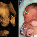
A flat profile (Fig. 4.4) associated with bilateral clefts with or without palate involvement has been described by Gabrielli et al. [10]. In their retrospective study with 14 fetuses with prenatal diagnosis of bilateral clefts, 9 of 14 fetuses had a premaxilla protuberance and 5 of 14 (approximately one-third) had a flat profile. The 5 fetuses with a flat profile were aneuploid, including three with trisomy 18, one with trisomy 13, and another with mosaic trisomy 8. In the group with premaxilla protuberance, only one of the nine fetuses had trisomy 13. The authors concluded that the flat profile is seen in approximately a third of the cases with prenatal diagnosis of bilateral clefting and that these fetuses are at high risk of having a lethal aneuploidy. The combination of a flat facial profile and a chromosomal abnormality suggests that many of these clefts, which are usually medial fissures, are a consequence of a deficiency during the development of the frontonasal process affecting the forehead and nose.
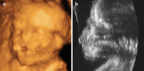

Fig. 4.4
(a) Surface rendered image using 3D ultrasound of a 16-week fetus affected with trisomy of chromosome 13 with a midline cleft. (b) Sagittal plane (2D ultrasound) of the same fetus illustrating the “flat profile”
In the anterior coronal plane, in which the tip of the nose and the length of the lip are seen in the same view, the interruption of the surface of the lip can be visualized and classified as unilateral (right or left) or bilateral (Fig. 4.5).
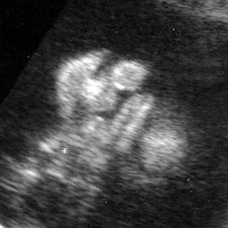

Fig. 4.5
Anterior coronal plane (2D ultrasound) of a bilateral CLP
In the transverse or axial plane performed with 2D ultrasound, we can identify the alveolar crest of the upper maxilla (Fig. 4.6). However, we cannot see the secondary palate (hard or soft) because of shadowing from the surrounding alveolus and the presence of the tongue.
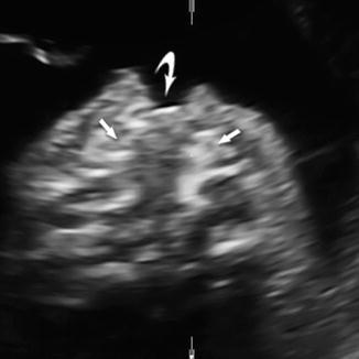

Fig. 4.6
Axial plane (2D ultrasound) of a left CLP showing the lip (curved arrow) and the affected border of the alveolus (straight arrows). Shadowing from the tongue and the ear precludes visualization of the secondary palate
In addition, evaluation of the uvula can be a helpful in determining if there may be a defect of the soft palate. Sonographic visualization of the uvula can be achieved by obtaining a coronal plane through the neck and pharynx or via a transverse plane with slight tilting of the transducer as demonstrated by Wilhelm and Borgers [11] in 667 consecutive patients with a normal singleton pregnancy between 20 and 25 weeks of gestation. These authors demonstrated that a normal uvula could be visualized with a typical echo pattern (the “equals sign”) in 90.7 % of the cases and the soft palate could be completely visualized in a median sagittal section in 85.3 % of the cases. Visualization of at least one of the two structures (either the uvula or the soft palate) was successful in 98.4 % of cases. Ultrasound detection of a bifid uvula (consisting in a cleft at this level) is diagnostic of a soft palatal defect [12].
In addition to the use of conventional 2D ultrasound, color Doppler ultrasound may contribute to the prenatal diagnosis of CP by demonstrating abnormal amniotic fluid flow between the oropharynx and the nasopharynx cavity, either using 2D or 3D ultrasound [13–15]. However, power Doppler may create a “blooming artifact” or “color bleed” [16] compared to high-definition power Doppler. The new bidirectional power Doppler technique combines high axial resolution with reduced spatial overlap of tissue and flow signals [17].
4.3 Exploration with 3D ultrasound
As we have highlighted above, in the second and third trimesters, defects involving the lips and the surrounding alveolus are often successfully diagnosed with relative ease using conventional 2D ultrasound. However, the diagnosis of the secondary palate involvement (hard and soft) continues to be a challenge for the sonographer.
With the arrival of 3D ultrasound, a series of techniques have been developed for exploring the secondary palate. These are the “reverse face” technique published by Campbell et al. [18], the “flipped face” technique developed by Platt et al. [19], and variations of both techniques [11, 20–24]. All seek to evaluate the palate in coronal or axial planes using a multiplanar system and/or rendering provided by 3D ultrasound. Our group published another method [25], which we termed the “oblique face” technique using the Oblique View tool that enables selection of nonconventional planes of volume (oblique or curved). The results are similar to that obtained with the OmniView tool [26].
To view the secondary palate, it is essential that the plane of initial acquisition of volume is very clear. It needs to be a midsagittal plane of the face, restricting the sector over the facial mass. According to the recommendations of Pilu and Segata [27], the fetus should have the head slightly deflected; the face may be gently pressed with the transducer to induce this slight deflection (Fig. 4.7) so that the hard palate and the shadow of the maxilla do not interfere with the ultrasound. We use maximum quality, high harmonics, and an angle of 40–70° depending on gestational age. It does not matter whether the cord or the placenta is in front of the face, but there should not be any limbs because of the shadows created over the upper maxilla or hard palate.
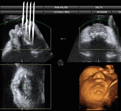

Fig. 4.7
Window A: The initial plane for volume acquisition is a straight midsagittal plane from which the volume is obtained with a sweep angle of 40–70°. The sweep is performed in a lateral direction from one side of the face to the other. The head is slightly deflected so that the ultrasound beams are directed toward the palate at a slightly oblique angle (arrows). Window B: coronal plane. Window C: axial plane. Window X: surface rendered image of the face
Below, we describe the techniques that enable us to visualize the secondary palate with 3D ultrasound.
4.3.1 “Reverse Face” View
According to the technique described by Campbell et al. [18], we place the green rendering lines from the posterior part to the anterior part of the palate. This allows coronal reconstructions rendered from the “inside” (Fig. 4.8).
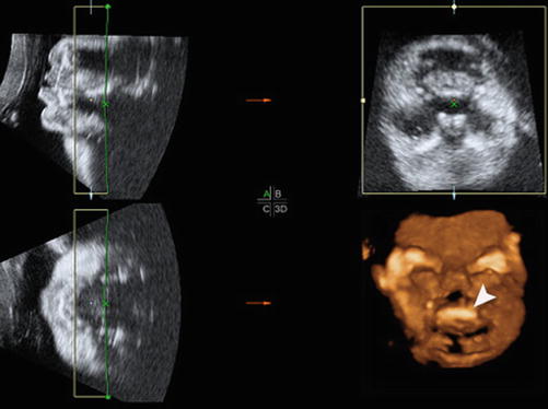

Fig. 4.8
Reverse face technique. Window A: initial plane of volume acquisition with the profile of the face turned 90° and the green rendering line placed to the right, from the inside of the face. Window B: coronal plane of the face. Window C: axial plane at the level of the nasal septum. Window X: surface rendered image demonstrating the hard palate in a coronal plane (arrow head)
4.3.2 “Flipped Face” View
This technique was described by Platt et al. [19]. We rotate the fetal face 90° from the midsagittal supine position to obtain axial planes of the secondary palate displaced from the chin up to the nose in a rendered surface reconstruction. We have enhanced this technique slightly; given that the form of the palate is concave, we curve the green line over the palate to achieve a better representation of the palate. The green line is curved over the coronal plane of the multiplanar evaluation (demonstrating the laterals borders of the palate) and the sagittal plane (demonstrating the anterior and posterior borders of the palate) (Fig. 4.9).
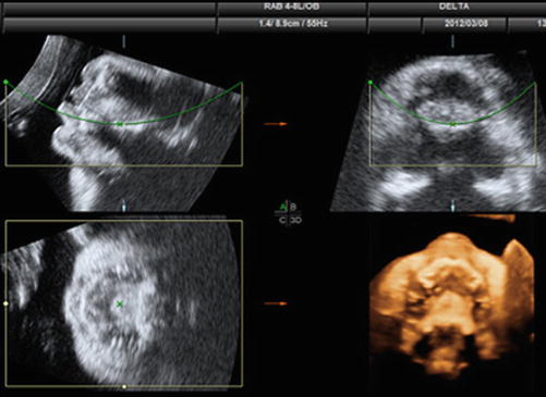

Fig. 4.9
Flipped face technique. Window A: initial plane of volume acquisition with the profile of the face turned 90° and the green line of the rendering box curved so that it is fitted to the location of the hard and soft palate. Window B: coronal plane of the face with the green line curved over the hard palate. Window C: axial plane at the level of the nasal septum. Window X: surface rendering on the right shows an axial plane of the maxilla with alveolar ridges, hard palate, and soft palate
4.3.3 “Oblique Face” View
To obtain the “oblique face” view, we use the Oblique View tool to obtain a nonstandard plane enhanced with XIMR [25] or the OmniView tool [26]. On a volume represented by the midsagittal section of the face, we specify a surface that traces the lips, the surrounding alveolus, and the palate, and we obtain an axial plane, perpendicular to this previously traced surface, in which the palate is incorporated. Secondly, we trace a line on the profile in the caudal-cranial direction from inside the face, and we obtain a coronal plane that we can shift and rotate along the length of the palate (Fig. 4.10).
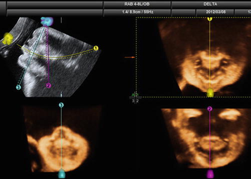

Fig. 4.10


Oblique face technique. We used the OmniView tool (GE Health Care, Zipf, Austria). Window A: initial plane of volume acquisition on which we trace a nonstandard plane (oblique or curved). The first line with volume contrast imaging (VCI) in the 2 mm thickness (1, yellow) is curved over the palate to include the upper lip and the central part of the upper maxilla of the hard and soft palate. In Window 1, the plane obtained perpendicular to the traced line is a curved axial plane that shows the lip, the upper maxilla with the alveolar holes, and the hard palate. The second line with VCI with a thickness of 2 mm (2, purple) is perpendicular to the hard palate. In Window 2, we see the obtained coronal plane that traverses the hard palate and is similar to that obtained with the Campbell (2007) technique. The third line with VCI in thickness of 2 mm is oblique (3, blue) and incorporates the more anterior part of the facial mass. In Window 3, we see an anterior coronal plane of the face with the central portion of the upper maxilla
Stay updated, free articles. Join our Telegram channel

Full access? Get Clinical Tree


