Fig. 3.1
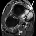
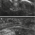
A 22-year-old male competitive downhill skier with thigh injury. Ultrasound images of the right thigh show a complex fluid collection over the right lateral thigh, containing layering fluid (arrows, a) and a nodule (arrow, b) which shows no increased color flow. Axial T2-weighted MRI (c) graphically demonstrates the relationship of the mass to the tensor fascia lata (TFL) and shows the nodule (arrow) within the fluid content. Coronal T1-weighted MRI (d) shows the bright T1 fat within the lesion (arrow) confirming the diagnosis of a Morel-Lavallee de-gloving injury (arrowheads delineate TFL, GT greater tuberosity, GM gluteus medius)
3.5 Conclusion
There is little doubt that participation in the imaging and care of athletes at major sporting events can be among the highlights of one’s career as a musculoskeletal radiologist or technologist. The team approach to diagnosis and intervention of not only athletes but also related personnel and spectators has become more evident, and the key role that medical imaging plays is clear. Careful planning of equipment, human, and information technology resources is critical to success. The motivation to be part of the imaging team is rooted in love of sport and competition (Collier 2012) and the realization that imaging can optimize an athlete’s performance and help them and their team to excel under often worldwide media scrutiny.
References
Bethapudi S, Robinson P, Engebretsen L, Budgett R, Vanhegan IS, O’Connor P (2013b) Elbow injuries at the London 2012 Summer Olympic games: demographics and pictorial imaging review. AJR Am J Roentgenol 201:535–549CrossRefPubMed
Stay updated, free articles. Join our Telegram channel

Full access? Get Clinical Tree


