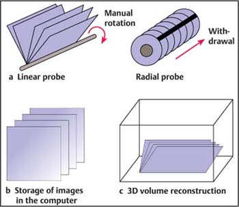
25 Three-Dimensional Endorectal Ultrasound and Rectal Cancer Several studies in the field of endoscopic ultrasound (EUS) technology have reported advantages with three-dimensional (3D) EUS.1–13 However, most 3D EUS studies have been conducted using catheter-type miniature probe systems.3,6–9 Some studies have previously reported benefits of a prototype 3D EUS system using a linear-array echoendoscope for 3D guidance of interventional procedures, but the scanning method used in this system has limitations and, as the ultrasound probe was not positioned at the tip of the endoscope, it was difficult to obtain clinically adequate images in the stomach without geometrical distortion.14–16 A recent study attempted to resolve this problem and maximize the performance of 3D EUS using a linear echoendoscope with a miniature electromagnetic position sensor attached to the tip of the scope, which can be used for freehand scanning in any position.17 However, the problem with this technique is that the electromagnetic sensor increases the size of the probe. This chapter reports on experience with a totally new 3D EUS software program that works without the need for an electromagnetic sensor and can be used even with electronic radial or linear rectal probes. Basic Principles of Three-Dimensional Ultrasound Two types of system have been developed, making use of either a series of two-dimensional (2D) images produced by one-dimensional arrays, or using 2D arrays to produce 3D images directly. Two criteria have to be met to avoid inaccuracies: the relative position and angulation of the acquired 2D images need to be known accurately; and the images have to be acquired rapidly and/or gated to avoid artifacts caused by respiratory, cardiac, and involuntary motion. Tracked free-hand systems. The operator holds an assembly consisting of the transducer and an attachment, and manipulates it over the anatomy. Two-dimensional images are digitized as the transducer is moved, while meeting two criteria: the precise relative angulation and position of the ultrasound transducer have to be known for each digitized image; and the operator has to ensure that no significant gaps are left when scanning the anatomy. Three-dimensional reconstruction. The 3D reconstruction process involves generating a 3D image from a digitized set of 2D images. The approach used was the voxel-based volume. The 2D images are built into a 3D voxel-based volume (3D grid) by placing each digitized 2D image into its correct location in the volume. The main advantages of this are that no information is lost during the 3D reconstruction and that a variety of rendering techniques are possible; however, large data files are generated. Visualization of three-dimensional ultrasound images. The ability to visualize information in the 3D image depends critically on the rendering technique. Three basic methods are used: • Surface-based viewing technique. An operator or algorithm identifies the boundaries of structures in order to create a wire-frame representation. These boundaries are shaded and illuminated, so that surfaces, structures, and organs are visualized. • Multiplane viewing techniques(Fig. 25.1). – Orthogonal views: three perpendicular planes are displayed simultaneously and can be moved or rotated. – Polyhedron: the 3D images are presented as a multiple-sided volume (polyhedron). The appropriate ultrasound image is “painted” onto each face of the polyhedron, which can be manipulated. • Volume-based rendering techniques. The 3D image is projected onto a 2D plane by casting rays through the 3D image. The voxel values intersected by each ray can be multiplied by factors and summed to produce different effects: multiplied by one and then added, to form a radiography-like image; multiplied by factors to produce translucency; or displaying only the voxel with the maximum intensity along each ray. Fig. 25.1a–c The various steps involved in three-dimensional endoscopic ultrasound (3D EUS) scanning using radial or linear echoendoscopes. Results of Three-Dimensional Endorectal Ultrasound in the Literature Our experience. From May 2003 to March 2004, we conducted 35 3D endorectal ultrasound (ERUS) examinations using this new software program. The indication for EUS was local staging of rectal cancer in all 35 cases. Before 3D ERUS scanning, a standard ERUS examination was performed. Three-dimensional rectal examinations were conducted using a radial electronic rigid probe (Hitachi R 54). The 3D software is included in the new ultrasound scanning machine (Hitachi 6500 or 8000) and allows reconstruction of the 2D EUS pictures in six different scans. The acquisition time is very fast, at around 10–25 seconds. The acquisition time was a function of the number of images recorded.
Related posts:

Stay updated, free articles. Join our Telegram channel

Full access? Get Clinical Tree


