Tibial Nerve Block

50 Tibial Nerve Block


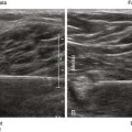
The tibial nerve is the largest branch of the sciatic nerve and the largest nerve for the ankle block. It provides sensory innervation to the heel and to the sole of the foot. The tibial nerve divides into the medial calcaneal, medial plantar, and lateral plantar branches near the ankle.1 In some subjects the takeoff of the medial calcaneal branch from the tibial nerve can be imaged above the ankle joint.


The order of anatomic structures from anterior to posterior at the medial malleolus is as follows: Tom, Dick, AVN, Harry (tibialis posterior tendon, flexor digitorum longus tendon, posterior tibial artery and veins, tibial nerve, flexor hallucis longus tendon). Therefore, the tibial nerve lies on the heel side of the posterior tibial artery. The posterior tibial artery is often accompanied by two flanking veins. This neurovascular bundle, consisting of one artery and two veins, can have a Mickey Mouse ears appearance if light touch with the transducer is applied (similar to the appearance of the brachial artery and veins near the elbow).


Edema or infection often makes routine ankle block ineffective or contraindicated.2 However, tibial nerve imaging can be difficult in some surgical patients with peripheral vascular disease because vascular landmarks for the nerve are not present. Tibial nerve block in the leg avoids the footdrop that occurs with more proximal popliteal block of the sciatic nerve. This can be an advantage for ambulatory surgery patients.


Suggested Technique


The tibial nerve can be approached in-plane from the posterior (Achilles) or anterior (tibial) side in supine position with the leg externally rotated using a short-axis view of the neurovascular bundle. The best point of tibial nerve imaging in the leg is usually halfway between the medial malleolus and the bulk of the gastrocnemius-soleus muscle complex in the calf. Place the block needle tip between the posterior tibial artery and the tibial nerve so as to enter the neurovascular compartment. With the posterior approach the Achilles tendon can lie close to the point of needle entry. With the anterior approach the saphenous vein can be close to the needle path near the skin surface.


Key Points


Only gold members can continue reading. Log In or Register to continue

Stay updated, free articles. Join our Telegram channel

Mar 5, 2016 | Posted by in ULTRASONOGRAPHY | Comments Off on Tibial Nerve Block

Full access? Get Clinical Tree

Get Clinical Tree app for offline access
Tibial Nerve Block The Essentials
Anatomy Two veins usually flank either side of the PTA.