(1)
Department of Radiology, John H Stroger Jr Hospital of Cook County, Chicago, IL, USA
Aorta
Diagnosis
Complete Aortic Rupture
Imaging Features
1.
Periaortic hematoma in direct continuity with the aortic wall
2.
Rupture at isthmus (commonest site)
3.
Intimal flap, traumatic pseudoaneurysm, intramural thrombus, abnormal aortic contour, and active extravasation of contrast
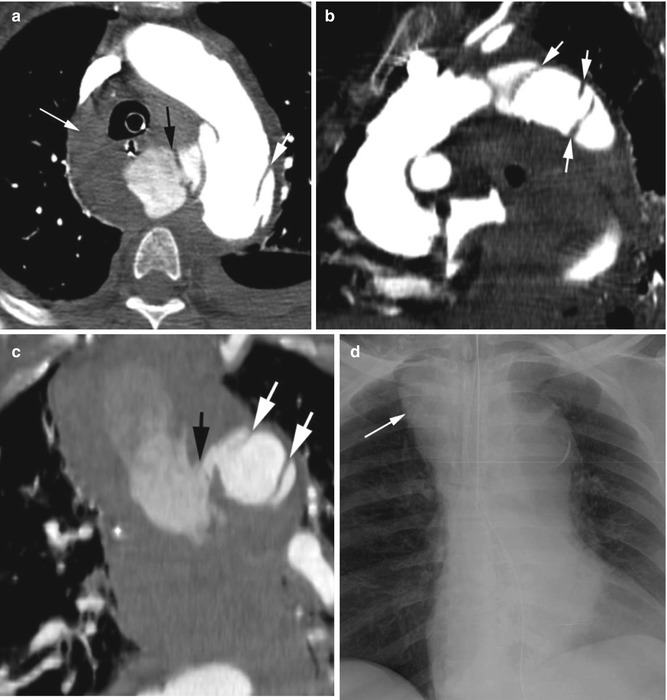
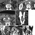
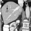
Fig. 4.1
Aortic transection. (a) Axial, (b) oblique sagittal, and (c) coronal reformatted CECT shows aortic transection at the isthmus (thick white arrows) with active hemorrhage into the mediastinum (black arrow), large hemomediastinum (long thin arrow) and left hemothorax. Multiple intimal flaps are seen (short white arrow). (d) Frontal chest X-ray shows hematoma widening the superior mediastinum (arrow)
Diagnosis
Incomplete Aortic Rupture
Imaging Features
1.
Saccular outpouching of the aortic lumen at anteromedial aspect of the isthmus by a collar
2.
Surrounding hemomediastinum
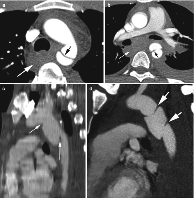
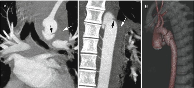
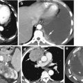
Fig. 4.2
Traumatic pseudoaneurysm of the aorta. (a, b) Axial CECT shows upper and lower end of the pseudoaneurysm at the isthmus (black arrows) with large hemomediastinum and hemothorax (white arrow). (c, d) Sagittal reformatted image in 2 patients shows pseudoaneurysm at the isthmus with dissection flaps at the neck (arrows). (e, f) Coronal reformatted images of patient in (d) show the upper and lower end of the tears (black arrows) with mediastinal hematoma (white arrow). (g) 3D volume-rendered image of the pseudoaneurysm. At surgery 90 % of the aorta was torn
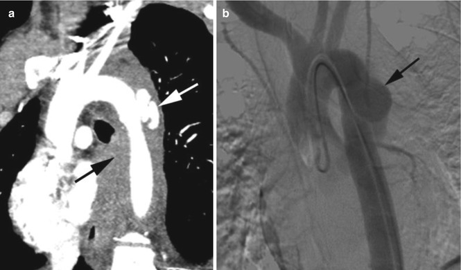
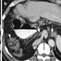
Fig. 4.3
Pseudoaneurysmic aorta following motor vehicle accident. (a) Sagittal reformatted image of lobulated saccular outpouching of pseudoaneurysm at anterolateral aortic isthmus (white arrow) with surrounding mediastinal hematoma (black arrow). (b) Angiogram showing pseudoaneurysm (arrow) with no extravasation of contrast
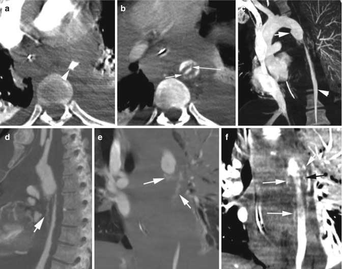
Fig. 4.4
Stanford type B dissection with circumferential intimomedial tear. High-speed MVA. Axial CT (a) early arterial phase shows narrowed contrast-filled descending thoracic aorta (arrow head). (b) Delayed arterial phase shows hematoma surrounding the true lumen with contrast filling the media at the periphery. Thin strands of contrast-filled intimal tear connecting to the medial tear (arrows). (c) Oblique coronal reformatted view shows pseudoaneurysm (arrow) at the aortic isthmus with constricted true lumen distal to it (arrow head). (d) Sagittal, (e, f) coronal reformatted images show irregular linear contrast filling the media from the pseudoaneurysm (arrows)
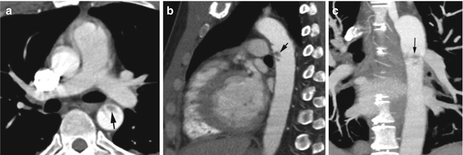
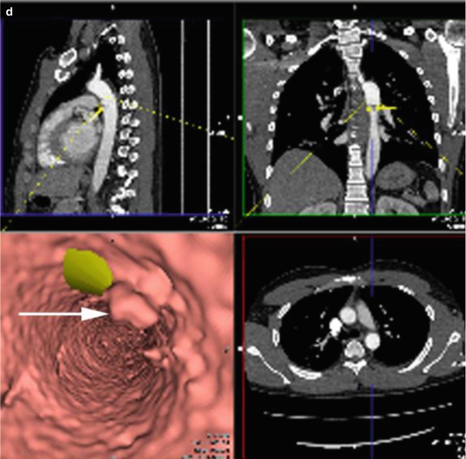
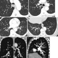
Fig. 4.5
Acute traumatic intramural hematoma at the aortic isthmus from deceleration MVA injury. (a) Axial, (b) sagittal, and (c) coronal reformatted images show irregular hematoma (arrow) in tunica media with no intimal flap and no significant mediastinal hematoma. (d) Flythrough endovascular postprocessed image shows protrusion of the intima into the lumen due to medial hematoma (arrow) with no intimal tear. The green patch represents the marking of arrow in the coronal and sagittal reformatted images
Pulmonary Artery
Diagnosis
Pseudoaneurysm of Left Pulmonary Artery
Imaging Features
1. Focal saccular outpouching of contrast in left main pulmonary arteryRelated posts:

Stay updated, free articles. Join our Telegram channel

Full access? Get Clinical Tree