3 Trigger Finger
♦ Setup
• The patient is seated facing the examiner with the hand resting supinated on the examination table.
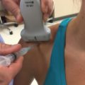
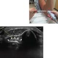
• Alternatively, the patient can be positioned supine with the hand supinated on a table or arm board facing the examiner (Fig. 3.1).
• A 12 to 21 MHz high-frequency linear probe with a footprint that is between 2.5 and 4.0 cm is used. A longer probe gives a longer field of view but tends to exhibit signal drop-off areas because of the varied contours of the surface of the hand.
♦ Landmarks
Several landmarks should be noted:
• Proximal finger crease (PFC)
• Valley of carpal tunnel

Fig. 3.1 The patient is positioned supine with the hand supinated toward the examiner. The proximal finger crease and valley of the carpal tunnel are located.
♦ Probe Positioning for Diagnostic Scan
Axial
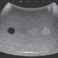
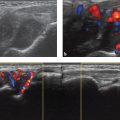
The probe should be placed 90 degrees to the long axis of the tendon sheath (Fig. 3.2).

Fig. 3.2 (a) The probe is placed 90 degrees to the long axis of the tendon sheath, approximately 1 cm proximal to the proximal finger crease. (b)The metacarpal head, proximal phalanx, flexor tendons, A1 pulley, and neurovascular bundles are visible in this axial direction.
Longitudinal
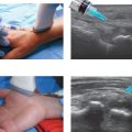
• The probe should be 90 degrees to the long axis of the tendon sheath at the level of the metacarpophalangeal joint (Fig. 3.3).
• The probe is centered proximal to the proximal finger crease to locate the A1 pulley.
• The probe should be perpendicular to the plane of the palm.
• The probe should be in line with the valley of the carpal tunnel.
• The probe should then be centered just over the proximal finger crease.
• The probe should be perpendicular to the floor and the plane of the palm.