Chapter 25 Tumours of the thorax
Lung cancer
Lung cancer remains the second most common cancer and is fatal in 85–90% of cases. This was a rare disease at the beginning of the twentieth century but, due to the explosion of smoking around the World War II, the incidence dramatically increased. By 1950, 80% of men and 40% of women smoked. During the early 1960s, the danger of tobacco was identified and, since the 1970s, the rates of smoking in men have reduced.
Unfortunately, due to a lag period until the development of cancer, the incidence of lung cancer today reflects smoking habits of the population 20 years previously. The incidence of lung cancer in men has been falling since 1990 but this is not the case in eastern European countries, in undeveloped countries or in women. Unfortunately, the incidence of female lung cancer is expected to increase until 2015 when it will almost equal rates in men.
Cigarette smoking accounts for 85–90% of cases. However, only 10–15% of smokers eventually develop lung cancer. There is probably interplay between genetic susceptibility of the disease and environmental factors such as air pollution and radon.
Pathology
Non-small cell lung cancer accounts for the majority of cases; 35–70% are squamous; 9–29% adenocarcinoma and 3–16% large cell. The incidence of small cell is decreasing and is currently 15–20% of tumours. Unfortunately, mesothelioma, which was previously rare, is rapidly increasing in incidence.
Symptoms
Primary
Ninety percent of patients are symptomatic on presentation. Central tumours produce symptoms of cough, pain and hemoptysis. Obstructive infective symptoms or lobar collapse may cause dysponea. If the mediastinum is directly invaded or mediastinal glands are present, hoarseness (secondary to recurrent laryngeal nerve involvement), dysphagia, superior vena caval obstruction and pericardial irritation or effusion develop.
Peripheral tumours may grow to a larger size before causing symptoms. Dysponea may be the presenting sign of a pleural effusion. Direct extension into the rib or brachial plexus causes Pancoast’s syndrome. This results in pain or sensory loss of the C7/8 T1 dermatomes and wasting of the small muscles of the hand. If the sympathetic plexus, which lies on the carotid artery, is affected Horner’s syndrome is also seen. This is characterized by miosis, ptosis, enophthalmus and ipsilateral anhydrosis.
Secondary
Invasion of the lymphatic system often occurs early. Hilar, mediastinal, neck and even axillary lymphadenopathy is possible. Once the basement epithelium is broken, the patient is at risk of hematogenous spread. Secondary involvement of the liver, adrenals, brain and bones is common.
Paraneoplastic syndromes
Tumours may produce proteins or hormones such as excess adrenocorticotrophic hormone (ACTH) leading to Cushing’s syndrome. Depending on the functionality of the protein, endocrine effects may be seen. Antigens expressed by the tumour may lead to cross tissue reactivity and syndromes suggestive of autoimmune disease.
Diagnosis
Patients presenting with the above symptoms require a chest x-ray. A lateral view may be helpful. A computed tomography (CT) scan of the chest and upper abdomen is recommended before bronchoscopy as peripheral tumours will not be reached by bronchoscopy and, in these cases, a CT-guided biopsy is required for histological diagnosis. Magnetic resonance imaging (MRI) can be used to determine if there are direct invasion structures contraindicating surgery but is not superior to CT in the detection of mediastinal disease (Table 25.1).
Table 25.1 Assessment of patients with lung cancer
History and examination including performance status and weight lossFBC, U&E, glucose, calcium, LFTChest x-ray and lateralBronchoscopy for histological/cytological diagnosis and to assess extent of the lesionCT of chest and upper abdomenAdditional tests are required depending on symptoms and the results of the above Additional staging if considering surgery: |
PET scanning
Positron emission tomography (PET scanning) is not currently widely used in the diagnosis and follow up of patients with lung cancer. The basis of PET scanning is the increased metabolic activity of cancer which avidly takes up glucose. Following administration of 18 fluorodeoxy-D-glucose (18F-FDG) there is emission of positrons from tumour-bearing areas. PET scanning is a high resolution, whole body technique which can demonstrate the extent of tumour spread. It is also useful in differentiating between benign and malignant pulmonary nodules. PET can detect lymph node spread more accurately than even spiral CT scanning.
One disadvantage of PET scanning is the rather limited anatomical information provided and a supplementary CT scan must be performed with image overlay to relate increased metabolic activity to anatomical sites. Moreover, it is possible to have a false positive scan.
Increased glucose metabolism may also be seen in a variety of inflammatory conditions including active tuberculosis, sarcoidosis abscesses and pneumonia.
Non-small cell lung cancer (NSCLC)
Staging
The NSCLC Staging system (Table 25.2) is based on the TNM (tumour, node, metastases) method of staging and divides patients into prognostic groups for treatment.
Table 25.2 The staging system of NSCLC
T stage | Disease extent |
---|---|
T0 | No evidence of tumour |
Tis | Carcinoma in situ |
Tx | Tumour cells obtained from sputum, no site of primary found on bronchoscopy |
T1 | Tumour <3 cm surrounded by visceral pleura or lung tissue |
T2 | Tumour larger than 3 cm but more than 2 cm from the carina. Involves the visceral pleura or causes atelectasis or obstructive pneumonitis of less than one lung |
T3 | Tumour less than 2 cm from the carina but not involving it. Tumour invades chest wall; diaphragm; mediastinal pleura; parietal pericardium or causes atelectasis or obstructive pneumonitis of the whole lung |
T4 | Tumour invades heart; great vessels; trachea; oesophagus; vertebral body; pleural effusion or satellite tumour nodules within the same lobe |
Nx | The nodal status cannot be determined |
N0 | No regional lymph node involvement |
N1 | Ipsilateral peribronchial and/or hilar lymphnode involvement |
N2 | Ipsilateral mediastinal and/or subcarinal lymphnodes |
N3 | Contralateral mediastinal or hilar lymphnodes. Supraclavicular lymphnodes |
Mx | Distant metastases cannot be determined |
M0 | Distant metastases absent |
M1 | Distant metastases present |
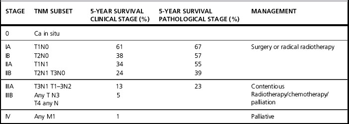
The staging and survival of NSCLC (Table 25.3) illustrates the prognostic importance of the current staging system.
The management of NSCLC
Surgery
Surgery offers the best chance of cure. Patients must be carefully selected, as incomplete excision is of no benefit. Patients must undergo extensive staging and, in practice, only those with stage I and II tumours and good cardiopulmonary function are suitable for surgery (Table 25.4).
Table 25.4 Contraindications for surgery in NSCLC
Operable | Contraindications |
---|---|
FEV1 > 1.5Stage I and II NSCLC | Poor lung functionPhrenic nerve palsy (elevated diaphragm)Recurrent laryngeal nerve palsy (hoarseness)Invasion of trachea, aorta, heart, superior vena cava, oesophagusDistant metastasesMetastatic SCLCMalignant pleural effusion |
Occasionally, patients may have surgically resectable disease but are unable to tolerate a surgical procedure due to medical co-morbidity. These patients may be considered for radical radiotherapy.
Radical radiotherapy
Thoracic radiotherapy is particularly challenging as the tumour is a moving target within an area surrounded by critical and radiosensitive tissues, such as lung parenchyma and spinal cord. In addition, the characteristics of the beam are altered as the beam passes through lung tissue. Careful planning is required to take these challenges into account (Table 25.5).
Table 25.5 Indications for radical radiotherapy
Stage 1&2 NSCLC – medically inoperableTumour volume approximately 5 cmFEV>1; FVC>1.5Weight loss <10%Positive margins postoperativelyHeavy N2 disease postoperatively (individual patient basis)Wedge resectionStage 3 disease sufficiently downstaged to be included in a radical volume |
Technique
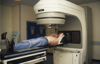
Patients should be CT planned, lying supine in an immobilization device. The patient is instructed to breathe normally. The normal treatment position is ‘arms up, elbows flexed’ but, for apical tumours, it may be better to have the arms down (Figure 25.1).
A CT scan is performed from the level of the larynx to the bottom of L2. This is to enable accurate delineation of the target and the lungs, spinal cord and other critical structures so that dose calculations to these structures are possible.
Target volume
Traditionally, ‘elective nodal irradiation’ was suggested for early stage NSCLC. Using this technique, the field extends from 5 to 8 cm below the carina, includes the entire mediastinum and bilateral hilar and extends superiorly to include the bilateral supraclavicular fossae. Such fields result in significant toxicity that limits the total dose deliverable. There is no evidence that this technique is superior to ‘involved field only’ techniques. The regional nodal recurrence with both is low, local recurrence more likely and the adoption of ‘involved field only’ treatment allows the possibility of dose escalation.
The ‘involved field only’ technique requires the treatment to be CT planned. The gross tumour volume (GTV) should be defined as all radiologically demonstrable tumour and any nodes over 1 cm in size. A planning target volume of GTV plus 1.5 cm in the lateral dimension and 2 cm vertical margin should be applied.
The type of plan created depends on the site of the tumour, whether central or peripheral. If the patient has undergone pneumonectomy, surgical clips may define the clinical target volume. It is important to ensure that beams pass through the resected space and spare the remaining contralateral lung (Figure evolves 25.2 and 25.3 ).
Usually, three fields are required for optimum dose distribution but, occasionally, if there is a small peripheral tumour, wedged opposed oblique fields may be used (Figure evolve 25.4 ).
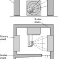
Depth dose tables estimating the dose at each point in tissue have been derived from experiments in solid tissue. Since air is much less dense, there is less scatter of the beam on traversing lung. As a result, the dose is 3% greater for every centimetre of lung traversed. Therefore, on calculating the dose to the tumour, the lung correction factor must be applied (Figure evolves 25.5 and 25.6 ).
Tolerances
It is hard to specify patient factors that predict the development of pneumonitis, but the mean lung dose and V20 (the volume of both lungs receiving more than 20 Gy) can help identify those particularly at risk.
A mean lung dose of 18–21 Gy is thought to give a low risk of development of pneumonitis, whereas 24–26 Gy patients are more likely to develop acute pneumonitis. V20, the volume of both lungs receiving more than 20 Gy, is probably the most reliable predictor at present and it is suggested that this value be less than 32% to avoid grade 3 or more pneumonitis.
The spinal cord should receive no more than 40–44 Gy depending on fraction size. Twelve centimeters of oesophagus should receive no more than 50 Gy. Heart doses at present are hard to define. They may not be reported routinely but the whole heart should receive less than 40 Gy and two thirds of it less than 55 Gy (Figure evolve 25.7 ).
Doses
Suggested radiation doses are given in Table 25.6.
Table 25.6 Suggested radiation doses for NSCLC
Indication | Suggested dose regimens |
---|---|
Radical postoperative dose | 50 Gy 20–23 fractions given in 4–5 weeks |
Radical radiotherapy | 55 Gy 20 fractions given in 4 weeks60 Gy 30 fractions given in 6 weeks |
Acute reactions
General fatigue and oesophagitis are common. Oeophagitis should respond to simple analgesics and antacids and should settle within 4 weeks of treatment.
Late reactions
Acute pneumonitis occurs in 10% of cases and is the most common and serious complication of radical treatment. Patients present with dry cough, fever and shortness of breath. A chest x-ray may show a hazy appearance, usually in the treated area. Therapy should be oxygen, antibiotics and high dose steroids (prednisolone 60 mg per day). Occasionally Lhermitte’s sign (an electric shock sensation when flexing the neck) is seen. Pulmonary fibrosis, esophageal stricture and pericardial effusion are seen in the longer term.

Full access? Get Clinical Tree

