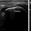
Fig. 14.1
The ulnar collateral ligament of the metacarpophalangeal joint of the thumb originates on the dorsal aspect of the head of the first metacarpal and continues on toward the volar aspect, where it inserts on the tubercle of the proximal phalanx (Fl p). The ligament lies deep to the aponeurosis of the adductor pollicis (Ap add), fibers of which insert into the tendon of the extensor pollicis longus (Est lp)
Skier’s thumb—a term often used inappropriately as a synonym for gamekeeper’s thumb [6]—is caused by acute traumatic radial hyperabduction of the thumb. This type of trauma can produce sprains (stretching of the ligament without disrupting the structural integrity of the fibers), partial or complete tears (Figs. 14.2 and 14.3, respectively), or the so-called Stener lesion (Fig. 14.4) [7]. Gamekeeper’s thumb is the result of chronic microtrauma that renders the ligament lax and nonfunctional [6].
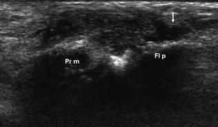
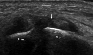
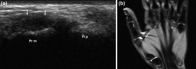
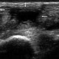
Fig. 14.2
Acute, partial-thickness tear of the ulnar collateral ligament of the metacarpophalangeal joint of the thumb. The ligament appears enlarged and hypoechogenic. In the distal portion, partial loss of continuity is evident with a hematoma between the ruptured fibers (arrow)
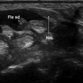
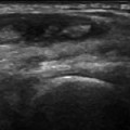
Fig. 14.3
Acute full-thickness tear of the ulnar collateral ligament of the metacarpophalangeal joint of the thumb. The ligament appears enlarged and hypoechogenic. Complete loss of continuity is evident and a hematoma can be seen between the stumps (arrow)
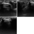
Fig. 14.4
Full-thickness tear of the ulnar collateral ligament of the metacarpophalangeal joint of the thumb with retraction of the proximal stump. The enlarged and hypoechogenic ligament stump is rolled back over the first metacarpal (Pr m) (arrows) (a, b), and there is an obvious joint effusion (b)
On ultrasound imaging, the ulnar collateral ligament is depicted as a hypoechoic structure situated on the ulnar side of the joint, between the head of the metacarpal and the proximal phalanx. It is covered by the hyperechoic aponeurosis of the adductor [7, 8]. The latter structure can be easily identified by having the patient flex his/her thumb. Because the aponeurosis inserts into the extensor pollicis longus, it also moves when the thumb is flexed [8].
In the presence of a sprain, sonographic examination will reveal hypoechogenicity of the injured segment but no loss of continuity.
Related posts:
Stay updated, free articles. Join our Telegram channel

Full access? Get Clinical Tree


